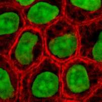
Cytokeratin
Cytokeratins are proteins of keratin-containing intermediate filaments found in the intracytoplasmic cytoskeleton of epithelial tissue. The term "cytokeratin" began to be used in the late 1970s (for example, see "Intermediate-sized filaments of human endothelial cells" by Franke, Schmid, Osborn and Weber[1]) when the protein subunits of keratin intermediate filaments inside cells were first being identified and characterized. In 2006 a new systematic nomenclature for keratins was created and now the proteins previously called "cytokeratins" are simply called keratins.[2]
Types

There are two types of cytokeratins: the acidic type I cytokeratins and the basic or neutral type II cytokeratins. Cytokeratins are usually found in pairs comprising a type I cytokeratin and a type II cytokeratin. Basic or neutral cytokeratins include CK1, CK2, CK3, CK4, CK5, CK6, CK7, and CK8. Acidic cytokeratins are CK9, CK10, CK12, CK13, CK14, CK16, CK17, CK18, CK19 and CK20. The cytokeratins can be divided into low versus high molecular weight solely based on their molecular weight. Expression of these cytokeratins is frequently organ or tissue specific. As an example, CK7 is typically expressed in the ductal epithelium of the genitourinary (GU) tract and CK20 most commonly in the gastrointestinal (GI) tract.[3] Anatomic pathologists employ such distinctions to detect the cell of origin of various tumors.
The subsets of cytokeratins which an epithelial cell expresses depends mainly on the type of epithelium, the moment in the course of terminal differentiation and the stage of development. Thus this specific cytokeratin fingerprint allows the classification of all epithelia upon their cytokeratin expression profile. Furthermore, this applies also to the malignant counterparts of the epithelia (carcinomas), as the cytokeratin profile tends to remain constant when an epithelium undergoes malignant transformation. The main clinical implication is that the study of the cytokeratin profile by immunohistochemistry techniques is a tool of immense value widely used for tumor diagnosis and characterization in surgical pathology.[4] The detection of soluble fragments of CK19 can be performed by ELISA using CYFRA 21-1 assay.
| Cytokeratin | Sites |
|---|---|
| Cytokeratin 4 |
|
| Cytokeratin 7 |
|
| Cytokeratin 8 |
|
| Cytokeratin 10 |
|
| Cytokeratin 13 |
|
| Cytokeratin 14 |
|
| Cytokeratin 18 |
|
| Cytokeratin 19 |
Does not react with hepatocytes and hepatocellular carcinoma[5] |
| Cytokeratin 20 |
|
Molecular biology
The cytokeratins are encoded by a family encompassing 30 genes. Among them, 20 are epithelial genes and the remaining 10 are specific for trichocytes.
All cytokeratin chains are composed of a central α-helix-rich domain (with a 50-90% sequence identity among cytokeratins of the same type and around 30% between cytokeratins of different type) with non-α-helical N- and C-terminal domains. The α-helical domain has 310-150 amino acids and comprises four segments in which a seven-residue pattern repeats. Into this repeated pattern, the first and fourth residues are hydrophobic and the charged residues show alternate positive and negative polarity, resulting in the polar residues being located on one side of the helix. This central domain of the chain provides the molecular alignment in the keratin structure and makes the chains form coiled dimers in solution.
The end-domain sequences of type I and II cytokeratin chains contain in both sides of the rod domain the subdomains V1 and V2, which have variable size and sequence. The type II also presents the conserved subdomains H1 and H2, encompassing 36 and 20 residues respectively. The subdomains V1 and V2 contain residues enriched by glycines and/or serines, the former providing the cytokeratin chain a strong insoluble character and facilitating the interaction with other molecules. These terminal domains are also important in the defining the function of the cytokeratin chain characteristic of a particular epithelial cell type.
Two dimers of cytokeratin groups into a keratin tetramer by anti-parallel binding. This cytokeratin tetramer is considered to be the main building block of the cytokeratin chain. By head-to-tail linking of the cytokeratin tetramers, the protofilaments are originated, which in turn intertwine in pairs to form protofibrils. Four protofibrils give place to one cytokeratin filament.

Cell biology
In the cytoplasm, the keratin filaments conform a complex network which extends from the surface of the nucleus to the cell membrane. Numerous accessory proteins are involved in the genesis and maintenance of such structure.
This association between the plasma membrane and the nuclear surface provides important implications for the organization of the cytoplasm and cellular communication mechanisms. Apart from the relatively static functions provided in terms of supporting the nucleus and providing tensile strength to the cell, the cytokeratin networks undergo rapid phosphate exchanges mediated depolymerization, with important implications in the more dynamic cellular processes such as mitosis and post-mitotic period, cell movement and differentiation.
Cytokeratins interact with desmosomes and hemidesmosomes, thus collaborating to cell-cell adhesion and basal cell-underlying connective tissue connection.
The intermediate filaments of the eukaryotic cytoskeleton, which the cytokeratins are one of its three components, have been probed to associate also with the ankyrin and spectrin complex protein network that underlies the cell membrane.
External links
- Cytokeratin at the US National Library of Medicine Medical Subject Headings (MeSH)
References
- ↑ Franke WW, Schmid E, Osborn M, Weber K (June 1979). "Intermediate-sized filaments of human endothelial cells". The Journal of Cell Biology. 81 (3): 570–80. doi:10.1083/jcb.81.3.570. PMC 2110384
 . PMID 379021.
 - ↑ Schweizer J, Bowden PE, Coulombe PA, et al. (July 2006). "New consensus nomenclature for mammalian keratins". The Journal of Cell Biology. 174 (2): 169–74. doi:10.1083/jcb.200603161. PMC 2064177. PMID 16831889.
- ↑ Levy, Gary; Purcel, Karen (2011). "Premalignant & Malignant Disorders of the Ovaries & Oviducts". In DeCherney, Alan; Nathan, Lauren; Goodwin, T. Murphy; Laufer, Neri; Roman, Ashley. Current Diagnosis & Treatment: Obstetrics & Gynecology. pp. 848–58. ISBN 978-0-07-163969-9.
- ↑ Walid MS, Osborne TJ, Robinson JS (2009). "Primary brain sarcoma or metastatic carcinoma?". Indian Journal of Cancer. 46 (2): 174–5. doi:10.4103/0019-509X.49160. PMID 19346656.
- 1 2 3 4 5 6 7 8 9 10 11 12 13 MUbio > MONOCLONAL ANTIBODIES TO CYTOKERATINS Retrieved October 2010
