T-tubule
| T-tubule | |
|---|---|
|
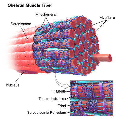
Skeletal muscle, with T-tubule labeled in zoomed in image. | |
|
Narrow T-tubules permit the conduction of electrical impulses. The SR functions to regulate intracellular levels of calcium. Two terminal cisternae (where enlarged SR connects to the T-tubule) and one T-tubule comprise a triad—a “threesome” of membranes, with those of SR on two sides and the T-tubule sandwiched in-between them. | |
| Details | |
| Identifiers | |
| Latin | tubulus transversus |
| Code |
TH H2.00.05.2.01018 TH H2.00.05.2.02013 |

A T-tubule (or transverse tubule) is a deep invagination of the sarcolemma, which is the plasma membrane of skeletal muscle and cardiac muscle cells. These invaginations allow depolarization of the membrane to quickly penetrate to the interior of the cell.
Structure
Each muscle fiber is surrounded by a sarcolemma (the muscle fiber's plasma membrane). Transverse tubules are invaginations of the sarcolemma. Though the invaginations are normally perpendicular to the length of the fiber, many T-tubules can lie axial to the long-axis of the fiber, both wrapping around and running alongside myofilament bundles. The size and arrangement of T-tubules vary depending on muscle type and species. T-tubules are thought to be rich in a number of proteins, including a large number of L-type calcium channels.
In skeletal muscle cells, T-tubules are typically located at the junction overlap between the A and I bands of the sarcomere, and together with a pair of terminal cisternae (bulbous enlarged areas of the sarcoplasmic reticulum) it forms an arrangement called a triad.
In cardiac muscle, the T-tubule membrane is normally opposed to a single terminal cisterna, which may wrap around the T-tubule. This occurs at the Z line and is called a diad.
It is physiologically important for excitation-contraction coupling (see section below) that the T-tubules are positioned close to the terminal cisternae of the sarcoplasmic reticulum, as the triad or diad arrangement allows physical and functional contact by voltage-dependent L-type calcium channels. So, an action potential along the sarcolemma causes calcium channels to open in the terminal cisternae/sarcoplasmic reticulum, which enables calcium to move from the sarcoplasmic reticulum into cytoplasm and the intracellular calcium concentration to increase.
Excitation-contraction coupling
See Excitation-contraction coupling
T-tubules are the major sites for the coupling of excitation and contraction, which is the process whereby the spreading depolarization is converted into force production by muscle fibers. The L-type calcium channels in T-tubules activate in response to electrical stimulation; their opening allows calcium to flow down its electrochemical gradient and into the cell. Activation of the L-type channel also causes a mechanical interaction between it and calcium-release channels located on the adjacent sarcoplasmic reticulum membrane.
In skeletal muscle, the influx of calcium through the L-type calcium channel on the T-tubule contributes little to excitation-contraction coupling, whereas it is crucial to the proper function of cardiac muscle (see Cardiac action potential). Conversely, the mechanical interaction between the T-tubule's L-type calcium channel and the calcium-release channel is critical to proper skeletal muscle contraction, whereas it contributes little to the contraction of cardiac muscle.
Detubulation
It is possible to physically and functionally uncouple T-tubules from the surface membrane using a technique known as detubulation. This relies on osmotically active chemicals, such as glycerol[1] (for skeletal muscle) or formamide (mainly for cardiac muscle). Addition of these chemicals to the solution surrounding muscle cells causes the cells to shrink; when the chemical is withdrawn the cells rapidly expand before returning to their normal size. The rapid expansion is thought to cause the t-tubules to detach from the surface membrane, which reseals, and to reseal within the cell. This technique can be used to investigate the function of the t-tubules.
There is evidence that heart failure precipitates the loss of the T-tubule network and this, in turn, contributes to the reduced contraction performance of the failing heart, underscoring their importance.[2]
See also
References
- ↑ Fraser, James A.; Skepper, Jeremy N.; Hockaday, Austin R.; Huang1, Christopher L.-H. (1998-08-01). "The tubular vacuolation process in amphibian skeletal muscle". Journal of Muscle Research & Cell Motility. 19 (6): 613–629. doi:10.1023/A:1005325013355. ISSN 0142-4319.
- ↑ Crossman DJ, Young AA, Ruygrok PN, Nason GP, Baddelely D, Soeller C, Cannell MB (May 2015). "t-tubule disease: Relationship between t-tubule organization and regional contractile performance in human dilated cardiomyopathy". Journal of Molecular and Cellular Cardiology. 84: 170–8. doi:10.1016/j.yjmcc.2015.04.022.
External links
- Histology image: 22502loa – Histology Learning System at Boston University - "Ultrastructure of the Cell: cardiac muscle, intercalated disk "
- Physiology: 2/2ch3/communic - Essentials of Human Physiology
