Pilonidal cyst
| Pilonidal cyst | |
|---|---|
 | |
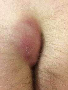
| Two pilonidal cysts that have formed in the gluteal cleft of an adult man. | |
| Classification and external resources | |
| Specialty | General surgery; Colorectal surgery |
| ICD-10 | L05 |
| ICD-9-CM | 685 |
| DiseasesDB | 31128 |
| eMedicine | emerg/771 |
| MeSH | D010864 |

Pilonidal cyst, also referred to as a pilonidal abscess, pilonidal sinus or sacrococcygeal fistula, is a cyst or abscess near or on the natal cleft of the buttocks that often contains hair and skin debris.[1]
Signs and symptoms
Pilonidal cysts are itchy and are often very painful, and typically occur between the ages of 15 and 35.[2] Although usually found near the coccyx, the condition can also affect the navel, armpit or genital region,[3] though these locations are much rarer.
Symptoms include:[4]
- Pain/discomfort or swelling above the anus or near the tailbone that comes and goes
- Opaque yellow (purulent) or bloody discharge from the tailbone area
- Unexpected moisture in the tailbone region
- Discomfort with sitting on the tailbone, doing sit-ups or riding a bike (any activities that roll over the tailbone area)
Some people with a pilonidal cyst will be asymptomatic.[5]
Pilonidal sinus
A sinus tract, or small channel, may originate from the source of infection and open to the surface of the skin. Material from the cyst may drain through the pilonidal sinus. A pilonidal cyst is usually painful, but with draining, the patient might not feel pain.
Causes
One proposed cause of pilonidal cysts is ingrown hair.[6] Excessive sitting is thought to predispose people to the condition, as sitting increases pressure on the coccygeal region. Trauma is not believed to cause a pilonidal cyst; however, such an event may result in inflammation of an existing cyst. However, there are cases where this can occur months after a localized injury to the area. Some researchers have proposed that pilonidal cysts may be the result of a congenital pilonidal dimple.[7] Excessive sweating can also contribute to the cause of a pilonidal cyst. Moisture can fill a stretched hair follicle, which helps create a low-oxygen environment that promotes the growth of anaerobic bacteria, often found in pilonidal cysts. The presence of bacteria and low oxygen levels hamper wound healing and exacerbate a forming pilonidal cyst.[8]
Differential diagnosis
A pilonidal cyst can resemble a dermoid cyst, a kind of teratoma (germ cell tumor). In particular, a pilonidal cyst in the gluteal cleft can resemble a sacrococcygeal teratoma. Correct diagnosis is important because all teratomas require complete surgical excision, if possible without any spillage, and consultation with an oncologist.
Treatment
Treatment may include antibiotic therapy, hot compresses and application of depilatory creams. In more severe cases, the cyst may need to be lanced or treated surgically. Lancing is performed using a local anesthesia, with healing time generally under one week.[9] The most conservative surgical treatment, Bascom's Pit Picking procedure, is a relatively simple outpatient option that can be performed in a physician's office, involves minimal pain and requires only a few days healing.[10] Although this procedure is much less invasive than the alternatives, it is not regularly practiced in the US. The Pit Picking procedure provides good results, fast recovery, and in instances where it is unsuccessful, other options for more invasive surgery can still be performed.
The more common course for surgical treatment is for the cyst to be surgically excised (along with pilonidal sinus tracts). Post-surgical wound packing may be necessary, and packing typically must be replaced once daily for 4 to 8 weeks. In some cases, two years may be required for complete granulation to occur. Sometimes the cyst is resolved via surgical marsupialization.[11]
Surgeons can also excise the sinus and repair with a reconstructive flap technique, such as a "cleft lift" procedure or Z-plasty, usually done under general anesthetic. This approach is especially useful for complicated or recurring pilonidal disease, leaves little scar tissue and flattens the region between the buttocks, reducing the risk of recurrence.[8] This approach typically results in a more rapid recovery than the traditional surgery, however there are fewer surgeons trained in the cleft lift procedure and thus, it may not be as accessible to patients, depending on their geographic location. Meta analysis shows recurrence rates were lower in open healing than with primary closure (RR 0.60, 95% CI 0.42 to 0.87) at the expense of time to healing.[12] Pilonidal cysts recur and do so more frequently if the surgical wound is sutured in the midline, as opposed to away from the midline, which obliterates the natal cleft and removes the focus of shearing stress. An incision lateral to the intergluteal cleft is therefore preferred, especially given the poor healing of midline incisions in this region.
An attractive minimally invasive technique is to treat pilonidal sinus with fibrin glue. This technique is less painful than traditional excisional techniques and flaps, can be performed under local or general anaesthesia, does not require dressings or packing and allows return to normal activities within 1 to 2 days. Long term outcome and recurrence rates are not dissimilar to more invasive techniques in 5 year follow up in a small randomised controlled trial.[13][14][15][16] Fibrin glue has also been shown to be better than more invasive alternatives in the treatment of pilonidal sinus disease in children, where a quick return to normal activities and minimal postoperative pain are especially important[17]
A minimally invasive surgical technique, was developed in Israel by Moshe Gips et al.,2008.[18] and is similar to the pit picking technique first described by Bascom in 1980 [19] In this procedure, trephines or biopsy punches which only "core out" and remove the diseased tissue and cyst are used, leaving only small holes to heal. Work or school activities will be resumed in one or two days, without or with minimal postoperative pain. The two procedures have been successfully combined by L. Basso in Rome (Italy).
While the recovery rate is positive for most, some suffer long term effects. Recorded instances include patients with continued postoperative pain for years following the surgical procedure. Primary complaints included pain when sitting for long period of times or following abrasive contact with the lower back and buttocks.
Etymology
Pilonidal means nest of hair and is derived from the Latin words for hair (pilus) and nest (nidus).[2] The condition was first described by Herbert Mayo in 1833.[20][21] R.M. Hodges was the first to use the phrase pilonidal cyst to describe the condition in 1880.[22][23]
The condition was widespread in the United States Army during World War II. The condition was termed "jeep seat" or "Jeep riders' disease", because a large portion of people who were being hospitalized for it rode in Jeeps, and prolonged rides in the bumpy vehicles were believed to have caused the condition due to irritation and pressure on the coccyx.
Notes
- ↑ Klass, Alan (November 1, 1956). "The So-Called Pilo-Nidal Sinus". Canadian Medical Association Journal. 75 (9): 737–742. PMC 1823328
 . PMID 13364825.
 - 1 2 "Pilonidal Cyst: Definition". Mayo Clinic. December 5, 2012. Retrieved February 8, 2013.
- ↑ Rao, Amrith; Sharma, Mohit; Thyveetil, Mabel; Karim, Omer (December 2006). "Penis: An Unusual Site for Pilonidal Sinus". International Urology and Nephrology. 38 (3–4): 607–608. doi:10.1007/s11255-005-4761-5. PMID 17111086.
- ↑ Sternberg, Jeffrey. "What Is Pilonidal Disease". Retrieved November 14, 2014.
- ↑ Doerr, Steven. "Pilonidal Cyst". eMedicineHealth. p. 1. Retrieved February 8, 2013.
- ↑ "Pilonidal Cyst: Causes". Mayo Clinic. December 5, 2012. Retrieved February 8, 2013.
- ↑ da Silva JH (2000). "Pilonidal cyst: cause and treatment". Dis. Colon Rectum. 43 (8): 1146–56. doi:10.1007/bf02236564. PMID 10950015.
- 1 2 Bascom, John; Bascom, Thomas (October 2002). "Failed Pilonidal Surgery". Archives of Surgery. 137 (10): 1146–50. doi:10.1001/archsurg.137.10.1146. PMID 12361421.
- ↑ "Pilonidal sinus disease". www.worldwidewounds.com. Retrieved 2016-05-22.
- ↑ Colov, Emilie Palmgren; Bertelsen, Claus Anders (2011-12-01). "Short convalescence and minimal pain after out-patient Bascom's pit-pick operation". Danish Medical Bulletin. 58 (12): A4348. ISSN 1603-9629. PMID 22142576.
- ↑ Prolonged delay in healing after surgical treatment of pilonidal sinus is avoidable
- ↑ AL-Khamis A, McCallumI, King PM, Bruce J. Healing by primary versus secondary intention after surgical treatment for pilonidal sinus. Cochrane Database of Systematic Reviews 2010, Issue 1. Art. No.: CD006213. DOI: 10.1002/14651858.CD006213.pub3.
- ↑ Elsey E, Lund JN (2013). "Fibrin glue in the treatment for pilonidal sinus: high patient satisfaction and rapid return to normal activities". Techniques in coloproctology. 17 (1): 101–104. doi:10.1007/s10151-012-0956-9.
- ↑ Lund JN, Leveson SH (2005). "Fibrin glue in the treatment of pilonidal sinus: results of a pilot study". Diseases of the Colon & Rectum. 48 (5): 1094–1096. doi:10.1007/s10350-004-0905-4.
- ↑ Liptrot S, Leveson S, Lund J (2008). "Fibrin glue may be better than surgery for pilonidal sinus: Results of a prospective, randomized, controlled trial and 2-year follow up". Diseases of the Colon & Rectum. 51 (5): 710–711.
- ↑ Isik A, Eryılmaz R, Okan I, Dasiran F, Firat D, Idiz O, Sahin M (Apr 2014). "The use of fibrin glue without surgery in the treatment of pilonidal sinus disease". Int J Clin Exp Med. 7 (4): 1047–51.
- ↑ Mary Smith Caroline; Jones Abigail; Dass Dipankar; Murthi Govind; Lindley Richard (2015). "Early experience of the use of fibrin sealant in the management of children with pilonidal sinus disease". Journal of Pediatric Surgery. 50 (2): 320–322. doi:10.1016/j.jpedsurg.2014.11.022.
- ↑ Gips M; et al. (2008). "Minimal surgery for pilonidal disease using trephines: description of a new technique and long-term outcomes in 1,358 patients". Dis Colon Rectum. 51: 1656–62. doi:10.1007/s10350-008-9329-x.
- ↑ Pilonidal disease: origin from follicles of hairs and results of follicle removal as treatment. Bascom J. Surgery. 1980. 87(5):567-72.
- ↑ Lanigan, Michael (September 27, 2012). "Pilonidal Cyst and Sinus". Medscape. WebMD. Retrieved February 8, 2013.
- ↑ Saad, Saad; Shakov, Emil; Sebastian, Vinod; Saad, Adam (2007). "The use of Wound Vacuum-assisted Closure (V.A.C.™) system in the treatment of Recurrent or Complex Pilonidal Cyst Disease: Experience in 4 Adolescent Patients". The Internet Journal of Surgery. 11 (1). doi:10.5580/382. ISSN 1528-8242.
- ↑ Hodges, RM (1880). "Pilonidal sinus". The Boston Medical and Surgical Journal. 103: 485–586.
- ↑ Kanerva 2000, p. 821
