Coccyx
| Coccyx | |
|---|---|
|
A coccyx with four vertebrae below the sacrum. | |
| Details | |
| Identifiers | |
| Latin | os coccygis |
| MeSH | A02.835.232.834.229 |
| TA | A02.2.06.001 |
| FMA | 20229 |

The coccyx (/ˈkɒksɪks/ KOK-siks; plural: coccyges or coccyxes), commonly referred to as the tailbone, is the final segment of the vertebral column in humans and apes, and certain other mammals such as horses. In animals with bony tails, it is known as tailhead or dock, in bird anatomy as tailfan. It comprises three to five separate or fused coccygeal vertebrae below the sacrum, attached to the sacrum by a fibrocartilaginous joint, the sacrococcygeal symphysis, which permits limited movement between the sacrum and the coccyx.
Structure
The coccyx is formed of either three, four or five rudimentary vertebrae. It articulates superiorly with the sacrum. In each of the first three segments may be traced a rudimentary body and articular and transverse processes; the last piece (sometimes the third) is a mere nodule of bone. The transverse processes are most prominent and noticeable on the first coccygeal segment. All the segments lack pedicles, laminae and spinous processes. The first is the largest; it resembles the lowest sacral vertebra, and often exists as a separate piece; the remaining ones diminish in size from above downward.
Most anatomy books incorrectly state that the coccyx is normally fused in adults. In fact it has been shown that the coccyx may consist of up to five separate bony segments, the most common configuration being two or three segments.[1][2]
Surfaces
The anterior surface is slightly concave and marked with three transverse grooves that indicate the junctions of the different segments. It gives attachment to the anterior sacrococcygeal ligament and the levatores ani and supports part of the rectum. The posterior surface is convex, marked by transverse grooves similar to those on the anterior surface, and presents on either side a linear row of tubercles–the rudimentary articular processes of the coccygeal vertebrae. Of these, the superior pair are the largest, and are called the coccygeal cornua they project upward, and articulate with the cornua of the sacrum, and on either side complete the foramen for the transmission of the posterior division of the fifth sacral nerve.
Borders
The lateral borders are thin and exhibit a series of small eminences, which represent the transverse processes of the coccygeal vertebrae. Of these, the first is the largest; it is flattened from before backward, and often ascends to join the lower part of the thin lateral edge of the sacrum, thus completing the foramen for the transmission of the anterior division of the fifth sacral nerve; the others diminish in size from above downward, and are often wanting. The borders of the coccyx are narrow, and give attachment on either side to the sacrotuberous and sacrospinous ligaments, to the coccygeus in front of the ligaments, and to the gluteus maximus behind them.
Apex
The apex is rounded, and has attached to it the tendon of the external anal sphincter. It may be bifid (divided into two).
Extensor coccygis
The extensor coccygis is a slender muscle fascicle, which is not always present. It extends over the lower part of the posterior surface of the sacrum and coccyx. It arises by tendinous fibers from the last segment of the sacrum, or first piece of the coccyx, and passes downward to be inserted into the lower part of the coccyx. It is a rudiment of the extensor muscle of the caudal vertebrae of other animals.
Sacrococcygeal and intercoccygeal joints
The joints are variable and may be: (1) synovial joints; (2) thin discs of fibrocartilage; (3) intermediate between these two; (4) ossified.[3][4]
Function
In humans and other tailless primates (e.g., great apes) since Nacholapithecus (a Miocene hominoid),[5][6] the coccyx is the remnant of a vestigial tail, but still not entirely useless;[7] it is an important attachment for various muscles, tendons and ligaments—which makes it necessary for physicians and patients to pay special attention to these attachments when considering surgical removal of the coccyx.[8] Additionally, it is also a part of the weight-bearing tripod structure which acts as a support for a sitting person. When a person sits leaning forward, the ischial tuberosities and inferior rami of the ischium take most of the weight, but as the sitting person leans backward, more weight is transferred to the coccyx.[8]
The anterior side of the coccyx serves for the attachment of a group of muscles important for many functions of the pelvic floor (i.e., defecation, continence, etc.): the levator ani muscle, which include coccygeus, iliococcygeus, and pubococcygeus. Through the anococcygeal raphe, the coccyx supports the position of the anus. Attached to the posterior side is gluteus maximus which extend the thigh during ambulation.[8]
Many important ligaments attach to the coccyx: the anterior and posterior sacrococcygeal ligaments are the continuations of the anterior and posterior longitudinal ligaments that stretches along the entire spine.[8] Additionally, the lateral sacrococcygeal ligaments complete the foramina for the last sacral nerve.[9] And, lastly, some fibers of the sacrospinous and sacrotuberous ligaments (arising from the spine of the ischium and the ischial tuberosity respectively) also attach to the coccyx.[8]
An extension of the pia mater, the filum terminale, extends from the apex of the conus, and inserts on the coccyx.
Clinical significance
Injuring the coccyx can give rise to a painful condition called coccydynia and one or more of the bones or the connections thereof may be broken, fractured tailbone.[10] [11] A number of tumors are known to involve the coccyx; of these, the most common is sacrococcygeal teratoma. Both coccydynia and coccygeal tumors may require surgical removal of the coccyx (coccygectomy). One very rare complication of coccygectomy is a type of perineal hernia known as a coccygeal hernia.[12]
History
Etymology
The term coccyx is derived from the ancient Greek word κόκκυξ[13][14] kokkyx "cuckoo";[15] the latter is attested in the writings of the Greek physician Herophilus to denote the end of the vertebral column.[16] This Greek name for the cuckoo was applied as the last three or four bones of the coccyx resemble the beak of this bird,[13][16][17][18] when viewed from the side.[8][19]
This established etymological explanation can also be found in the writings of the 16th century anatomist Andreas Vesalius whom wrote: os cuculi, a similitudine rostri cuculi avis [16] (the cuckoo bone shows a likeness to the beak of the cuckoo bird). Vesalius used the Latin expression os cuculi, with os, bone[20] and cuculus, the Latin name for the cuckoo.[20] The 16th/17th century French anatomist Jean Riolan the Younger gives a rather hilarious etymological explanation, as he writes: quia crepitus, qui per sedimentum exeunt, ad is os allisi, cuculi vocis similitudinem effingunt [16] (because the sound of the farts that leave the anus and dash against this bone, shows a likeness to the call of the cuckoo). The latter is not considered as potential candidate.[16][17]
Besides os cuculi, os caudae,[16][21] with caudae, of the tail[20] is attested. This Latin expression might be the source of the English, French language, German and Dutch terms tailbone, l'os de la queue,[21] Schwanzbein [17][21] and staartbeen.[22] In the current official anatomic Latin nomenclature, Terminologia Anatomica,[23] coccyx and os coccygis is used.
Additional images
-
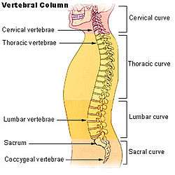
Vertebral column.
-
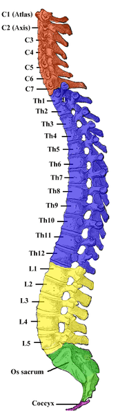
Vertebral column.
-

Left Levator ani from within.
-
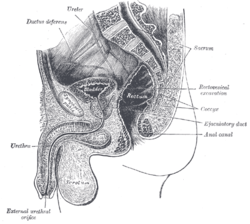
Median sagittal section of male pelvis.
-

Median sagittal section of female pelvis.
See also
| Wikimedia Commons has media related to Coccyx. |
References
- ↑ Postacchini F; Massobrio M] (October 1983). "Idiopathic coccygodynia. Analysis of 51 operative cases and a radiographic study of the normal coccyx". The Journal of bone and joint surgery (American volume ed.) (65(8)): 1116–1124.
- ↑ Kim NH, Suk KS (June 1999). "Clinical and radiological differences between traumatic and idiopathic coccygodynia". Yonsei Medical Journal (40:3): 215–220.
- ↑ Maigne JY; Molinie V; Fautrel B. (1992). "Anatomie des disques coccygiens". Revue de Médecine Orthopedique. 28: 34–35.
- ↑ Saluja PG. (1988). "The incidence of ossification of the sacrococcygeal joint". Journal of Anatomy. 156: 11–15.
- ↑ Nakatsukasa 2004, Acquisition of bipedalism (See Fig. 5 entitled First coccygeal/caudal vertebra in short-tailed or tailless primates..)
- ↑ Note: Nacholapithecus and Nakaliphitecus nakayamai are two different species of Miocene hominoids (specimens from Nakali and Nachola respectively). See for example "Comparisons with Other Hominoids" in (Kunimatsu, Nakatsukasa et al. Dec 2007)
- ↑ Saladin, Kenneth S. (2003). 3rd, ed. Anatomy & Physiology: The Unity of Form and Function. McGraw-Hill. p. 268.
- 1 2 3 4 5 6 Foye, Patrick M; Buttaci, Charles J (June 3, 2008). "Coccyx Pain". eMedicine.
- ↑ Morris, Craig E. (2005). Low Back Syndromes: Integrated Clinical Management. McGraw-Hill. p. 59. ISBN 0-07-137472-8.
- ↑ Maigne, J-Y; Doursounian, L; Chatellier, G.] (2000). "Causes and Mechanisms of Common Coccydynia. Spine". 25 (23). coccyx.org: 3072–3079.
- ↑ Foye P, Buttaci C, Stitik T, Yonclas P (2006). "Successful injection for coccyx pain.". Am J Phys Med Rehabil. 85 (9): 783–784. doi:10.1097/01.phm.0000233174.86070.63. PMID 16924191.
- ↑ Miranda EP, Anderson AL, Dosanjh AS, Lee CK (September 2007). "Successful management of recurrent coccygeal hernia with the de-epithelialised rectus abdominis musculocutaneous flap". J Plast Reconstr Aesthet Surg. 62 (1): 98–101. doi:10.1016/j.bjps.2007.08.002. PMID 17889632.
- 1 2 Klein, E. (1971). A comprehensive etymological dictionary of the English language. Dealing with the origin of words and their sense development thus illustration the history of civilization and culture. Amsterdam: Elsevier Science B.V.
- ↑ Harper, Douglas. "coccyx". Online Etymology Dictionary.
- ↑ κόκκυξ. Liddell, Henry George; Scott, Robert; A Greek–English Lexicon at the Perseus Project.
- 1 2 3 4 5 6 Hyrtl, J. (1880). Onomatologia Anatomica. Geschichte und Kritik der anatomischen Sprache der Gegenwart. Wien: Wilhelm Braumüller. K.K. Hof- und Universitätsbuchhändler.
- 1 2 3 Kraus, L.A. (1844). Kritisch-etymologisches medicinisches Lexikon (Dritte Auflage). Göttingen: Verlag der Deuerlich- und Dieterichschen Buchhandlung.
- ↑ Panourias, I.G., Stranjalis, G., Stavrinou, L.C. & Sakas, D.E. (2011). The Hellenic and Hippocratic origins of the spinal terminology. Journal of the History of the Neurosciences, 20, 177-187.
- ↑ Sugar, Oscar (February 1995). "Historical Perspective Coccyx: The Bone Named for a Bird". Spine. 20 (3): 379–383. doi:10.1097/00007632-199502000-00024. ISSN 0362-2436. PMID 7732478.
- 1 2 3 os, cuculus, cauda. Charlton T. Lewis and Charles Short. A Latin Dictionary on Perseus Project.
- 1 2 3 Schreger, C.H.Th.(1805). Synonymia anatomica. Synonymik der anatomischen Nomenclatur. Fürth: im Bureau für Literatur.
- ↑ Everdingen, J.J.E. van, Eerenbeemt, A.M.M. van den (2012). Pinkhof Geneeskundig woordenboek (12de druk). Houten: Bohn Stafleu Van Loghum.
- ↑ Federative Committee on Anatomical Terminology (FCAT) (1998). Terminologia Anatomica. Stuttgart: Thieme
- Kunimatsu, Yutaka; Nakatsukasa, Masato; et al. (December 2007). "A new Late Miocene great ape from Kenya and its implications for the origins of African great apes and humans". PNAS. 104 (49): 19220–5. doi:10.1073/pnas.0706190104. PMC 2148271
 . PMID 18024593. Retrieved January 2010. Check date values in:
 |access-date=(help)
- Nakatsukasa, Masato (May 2004). "Acquisition of bipedalism: the Miocene hominoid record and modern analogues for bipedal protohominids". Journal of Anatomy. 204 (204(5)): 385–402. doi:10.1111/j.0021-8782.2004.00290.x. PMC 1571308. PMID 15198702.
External links
- Anatomy photo:41:os-0107 at the SUNY Downstate Medical Center – "The Female Perineum: Osteology"
- Coccydynia (coccyx pain, tailbone pain) at eMedicine (Peer-reviewed medical chapter, available free online)