Nevus anemicus
| Nevus Anemicus | |
|---|---|
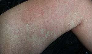 | |
| A man's left leg with nevus anemicus patches. | |
| Classification and external resources |
Nevus anemicus is a congenital disorder characterized by macules of varying size and shape that are paler than the surrounding skin and cannot be made red by trauma, cold, or heat.[1][2] The paler area is due to the blood vessels within the area which are more sensitive to the body’s normal vasoconstricting chemicals.[3]
Symptoms
This benign patch appears on the skin at birth or in early childhood. In most people these are under 10cm in size. If there is doubt about the diagnosis, rubbing the area causes the skin around the lesion to become red while the lesion itself does not change in color.[3] Often the patches are difficult to see against the background color of the patient’s skin, but if sunburn develops, then the white area stands out prominently.[4] The involved area is lighter than the normal skin, not because of a loss of pigment occurs, but because blood vessels are constricted, producing a permanent blanching of the area. This blanching is a functional rather than a structural abnormality, presumed to be caused by local increased sensitivity to catecholamines.[5] Although the cutaneous vasculature appears normal histologically, the blood vessels within the nevus do not respond to injection of vasodilators. It has been postulated that the persistent pallor may represent a sustained localized adrenergic vasoconstriction.[6] Results of a skin biopsy would be interpreted as normal and only physiological testing can reveal the nevus in contrast to normal skin. Stroking the patch elicits no red flare. Only the normal skin would react with a characteristic erythematous response. Examination under a Wood lamp can also reveal the nevus anemicus patch will not emphasize as a patch of vitiligo would.[7]
Treatment
Since the histopathology of nevus anemicus is normal, nevus anemicus is a pharmacologic nevus and not an anatomic one.[8][9] In most people a nevus anemicus is on a covered area and so light in appearance that no treatment is needed.[3]
See also
References
- ↑ James, William; Berger, Timothy; Elston, Dirk (2005). Andrews' Diseases of the Skin: Clinical Dermatology. (10th ed.). Saunders. Page 582. ISBN 0-7216-2921-0.
- ↑ Rapini, Ronald P.; Bolognia, Jean L.; Jorizzo, Joseph L. (2007). Dermatology: 2-Volume Set. St. Louis: Mosby. ISBN 1-4160-2999-0.
- 1 2 3 "Dermatologic Disease Database". Retrieved 25 October 2011.
- ↑ "Global Skin Atlas". Retrieved 25 October 2011.
- ↑ McMillan , Julia A; DeAngelis, Catherine D.; Feigin, Ralph D.; Warshaw, Joseph B. (2006). Oski's Pediatrics, Principles & Practice. (4th ed.). Page 458. ISBN 978-0-7817-1618-5.
- ↑ Nelson Textbook of Pediatrics 19th. (10th ed.). W.B. Saunders Company. Page 640. ISBN 1-4377-0755-6.
- ↑ Barber Kirk (2010). Consultant For Pediatricians '. Page 9:233, 237.
- ↑ Mountcastle EA, Diestelmeier MR, Lupton GP. Nevus anemicus. J Am Acad Dermatol 1986;14:628-32..
- ↑ Requena L, Sangueza OP. Cutaneous vascular anomalies. Part 1. Hamartomas, malformations and dilation of preexisting vessels. J Am Acad Dermatol 1997;37:523-49,quiz 549-52.
