Vitreous body
| Vitreous humour | |
|---|---|
 Schematic diagram of the human eye. | |
| Details | |
| Identifiers | |
| Latin | humor vitreus |
| TA | A15.2.06.014 |
| FMA | 67388 |
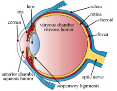
The vitreous body is the clear gel that fills the space between the lens and the retina of the eyeball of humans and other vertebrates. It is often referred to as the vitreous humour or simply "the vitreous".
Structure
The vitreous humour is a transparent, colorless, gelatinous mass that fills the space in the eye between the lens and the retina. It is present at birth.[1] Produced by cells in the non-pigmented portion of the ciliary body, the vitreous humor is derived from embryonic mesenchyme cells, which degenerate after birth.
The vitreous humour is in contact with the retina and helps to keep it in place by pressing it against the choroid. It does not adhere to the retina, except at the optic nerve disc and the ora serrata (where the retina ends anteriorly), at the Wieger-band, the dorsal side of the lens. It is not connected at the macula, the area of the retina which provides finer detail and central vision.
Significance
Clinical
Unlike the fluid in the frontal parts of the eye (aqueous humour) which is continuously replenished, the gel in the vitreous chamber is stagnant. Therefore, if blood, cells or other byproducts of inflammation get into the vitreous, they will remain there unless removed surgically. These are known as floaters. If the vitreous pulls away from the retina, it is known as a vitreous detachment. As the human body ages, the vitreous often liquefies and may collapse. This is more likely to occur, and occurs much earlier, in eyes that are nearsighted (myopia). It can also occur after injuries to the eye or inflammation in the eye (uveitis).
The collagen fibres of the vitreous are held apart by electrical charges. With aging, these charges tend to reduce, and the fibres may clump together. Similarly, the gel may liquefy, a condition known as synaeresis, allowing cells and other organic clusters to float freely within the vitreous humour. These allow floaters which are perceived in the visual field as spots or fibrous strands. Floaters are generally harmless, but the sudden onset of recurring floaters may signify a posterior vitreous detachment (PVD) or other diseases of the eye.
The metabolic exchange and equilibration between systemic circulation and vitreous humour is so slow that vitreous humour is sometimes the fluid of choice for postmortem analysis of glucose levels or substances which would be more rapidly diffused, degraded, excreted or metabolized from the general circulation.
Forensic
After death, the vitreous resists putrifaction longer than other body fluids. The vitreous potassium concentration rises so predictably within the hours, days and weeks after death, that vitreous potassium levels are frequently used to estimate the time-of-death (Post-mortem interval) of a corpse.[2][3][4]
Properties
Biochemical
Its composition is similar to that of the cornea, but the vitreous contains very few cells. It is composed mostly of phagocytes, which remove unwanted cellular debris in the visual field, and hyalocytes, which turn over the hyaluronan.
The vitreous humor contains no blood vessels, and 98-99% of its volume is water (as opposed to only 75% in the cornea). In addition to water, the vitreous consists of salts, sugars, vitrosin (a type of collagen), a network of collagen type II fibrils with glycosaminoglycan, hyaluronan, opticin, and a wide array of proteins. Despite having little solid matter, the fluid is substantial enough to fill the eye and give it its spherical shape. The lens, on the other hand, is tightly packed with cells.[5] The vitreous humour has a viscosity two to four times that of water, giving it a gelatinous consistency. It has a refractive index of 1.336.[6]
| Solute | Mean concentration | Units | Reference | Data from living humans? |
|---|---|---|---|---|
| Sodium | 146.7 | mmol/L | [7] | Yes |
| Potassium | 5.73 | mmol/L | [7] | Yes |
| Chloride | 121.6 | mmol/L | [7] | Yes |
| Calcium | 1.13 | mmol/L | [7] | Yes |
| Magnesium | 0.9 | mmol/L | [7] | Yes |
| Phosphate | 0.1 to 3.3 | mEq/dm3 | [8] | No |
| Bicarbonate | 1.2 to 3.0 | g/kg water | [8] | No |
| Solute | Mean concentration | Units | Reference | Data from living humans? |
|---|---|---|---|---|
| Copper | 0.52 | µmol/L | [7] | Yes |
| Selenium | 0.104 | µmol/L | [7] | Yes |
| Iron | 3.11 | µmol/L | [7] | Yes |
| Manganese | 110.7 | nmol/L | [9] | Yes |
| Solute | Mean concentration | Units | Reference | Data from living humans? |
|---|---|---|---|---|
| Glucose | 2.97 | mmol/L | [7] | Yes |
| Lactate | 3.97 | mmol/L | [7] | Yes |
| Lipids | 2 | μg/mL | [8] | No |
| Total protein content | 280-1360 | μg/cm3 | [8] | No |
| Hyaluronan | 42-400 | μg/cm3 | [8] | No |
| Versican | 60 | μg/cm3 | [8] | No |
| Collagen | 300 | μg/mL | [10] | No |
| Albumin | 293 ± 18 | μg/cm3 | [8] | No |
| Immunoglobulin (IgG) | 33.5 ± 3 | μg/cm3 | [8] | No |
| α1-Antitrypsin | 141 ± 2.9 | μg/cm3 | [8] | No |
| α1-Acid glycoprotein | 4 ± 0.7 | μg/cm3 | [8] | No |
| Osmolality | 289.5 | mOsm/kg | [7] | Yes |
| Beta-hydroxybutyrate | 0.094 | mmol/L | [7] | Yes |
| Ferritin | 19.52 | µg/L | [7] | Yes |
| Transferrin | 0.088 | g/L | [7] | Yes |
| Urea | 24-172 | mg/dL water | [8] | No |
| Creatinine | 0.3-3.0 | mg/dL water | [8] | No |
| Citrate | 1.9 | mg/dL water | [8] | No |
| Pyruvic acid | 7.3 | mg/dL water | [8] | No |
| Ascorbic acid | 36 | mg/100g | [8] | No |
Physical
| Property | Value | Units | Reference | Data from living humans? |
|---|---|---|---|---|
| Volume | 3.9 | mL | [8] | No |
| Weight | 3.9 | g | [8] | No |
| Water content | 99 to 99.7 | % | [8] | No |
| pH | 7.4 to 7.52 | [8] | No | |
| Osmolality | 289.5 | mOsm/kg | [7] | Yes |
| Osmotic pressure (Freezing-point depression) | -0.554 to -0.518 | °C | [8] | No |
| Density | 1.0053 to 1.0089 | g/cm3 | [8] | No |
| Intrinsic viscosity | 3-5 × 10³ | cm3/g | [8] | No |
| Dynamic viscosity | 1.6 | cP | [8] | No |
| Refractive index | 1.3345 to 1.337 | [8] | No |
| Age (years) | Gel volume (cm3) | Liquid volume (cm3) | Reference |
|---|---|---|---|
| Birth | 1.6 | 0 | [8] |
| 5 | 3.3 | 0 | [8] |
| 10 | 3.5 | 0.7 | [8] |
| 20 | 3.9 | 0.9 | [8] |
| 30 | 3.9 | 0.9 | [8] |
| 40 | 3.9 | 0.9 | [8] |
| 50 | 3.5 | 1.3 | [8] |
| 60 | 3.2 | 1.6 | [8] |
| 70 | 2.8 | 2.0 | [8] |
| 80 | 2.5 | 2.3 | [8] |
| 90 | 2.2 | 2.6 | [8] |
| Wavelength (nm) | Transmittance (total, %) | Reference | Data from living humans? |
|---|---|---|---|
| 300 | 0 | [11] | Yes |
| 325 | 76 | [11] | Yes |
| 350 | 82 | [11] | Yes |
| 400 | 90 | [11] | Yes |
| 500 | 97 | [11] | Yes |
| 600 | 98 | [11] | Yes |
| 700 | 98 | [11] | Yes |
| Parameter | Anterior vitreous | Central vitreous | Posterior vitreous | Units | Reference |
|---|---|---|---|---|---|
| Residual viscosity | 1.4 | 2.2 | 4.9 | ηm (Pa s) | [8] |
| Internal viscosity | 0.3 | 0.35 | 0.5 | ηk (Pa s) | [8] |
| Relaxation time | 0.38 | 0.30 | 1.61 | τm (s) | [8] |
| Retardation time | 0.27 | 0.41 | 0.46 | τk (s) | [8] |
| Elastic compliance (instantaneous) | 0.1 | 0.3 | 0.3 | Jm (m−2 N−1) | [8] |
| Elastic modulus (internal) | 2.5 | 1.3 | 1.2 | Gk (Pa) | [8] |
Anatomical
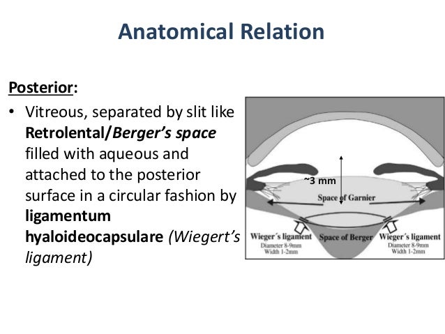
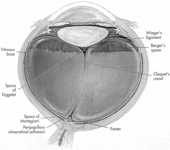
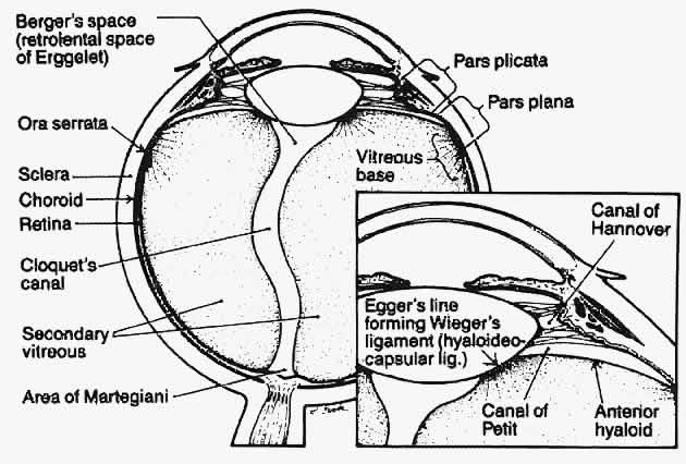
The vitreous has many anatomical landmarks, including the hyaloid membrane, Berger's space, space of Erggelet, Wieger's ligament, Cloquet's canal and the space of Martegiani.[12][13][14]
Surface features:
- Patella fossa: Shallow saucer-like concavity anteriorly, in which the lens rests, separated by Berger's space
- Ligamentum hyaloideocapsulare (Wieger's ligament): Circular thickening of vitreous 8-9mm in diameter, delineates the patella fossa
- Anterior hyaloid: Vitreous surface anterior to ora serrata. Continuous with and invests in the zonular fibres, and extends forward between the ciliary processes
- Vitreous base: Denser cortical area of vitreous. Firmly attached to the posterior 2mm of the pars plana, and the anterior 2-4mm of retina
- Posterior hyaloid surface: Closely applied to retinal internal limiting membrane. Firm attachment sites: Along blood vessels and at sites of retinal degeneration
- Space of Martegioni: A funnel shaped space overlying the optic disc with condensed edge
- Cloquet's canal: A 1-2mm wide canal within the vitreous, from the space of Martegioni to the space of Berger, along an S-shaped course mainly below the horizontal.
Internal structures of the vitreous
- The vitreous body at birth is homogenous with a finely striated pattern.
- With early aging the vitreous develops narrow transvitreal "channels".
- The cortex is denser than the centre with development.
- From adolescence, vitreous tracts form from anterior to posterior.
- These vitreous tracts are fine sheet-like condensations of vitreous.
Named tracts
- Retrolental tract: Extends posteriorly from the hyaloideocapsular ligament into central vitreous
- Coronary tract: External to the retrolental tract, and excluding posteriorly from a circular zone overlying the posterior 1/3rd of the ciliary processes
- Median tract: Extends back from a circular zone external to the coronary tract, at the anterior margin of the vitreous base
- Preretinal tract: Extends back from the ora serrata and vitreous base
Aging
- Birth: Homogenous and finely striated (except Cloquet's canal)
- Adolescence: the vitreous cortex becomes more dense and vitreous tracts develop
- Adulthood: Tracts become better defined and sinuous. Central vitreous liquefies, fibrillar degeneration occurs, and the tracts break up (syneresis)
- Aging: course strands. Cortex may disappear at sites, leading to liquid vitreous extruding into the potential space between vitreous cortex and retina (vitreous detachment)
- Posterior vitreous detachment: Once liquid vitreous enters the sub-hyaloid space between vitreous cortex and retina, it strips the vitreous cortex off the retina with each eye movement.
Additional images
-
Structures of the eye labeled
-

This image shows another labeled view of the structures of the eye
See also
References
- ↑ "Associated Structures - Vitreous". Teaching.pharmacy.umn.edu. Retrieved 2012-12-07.
- ↑ Zilg, B.; Bernard, S.; Alkass, K.; Berg, S.; Druid, H. (17 July 2015). "A new model for the estimation of time of death from vitreous potassium levels corrected for age and temperature". 254: 158–166. doi:10.1016/j.forsciint.2015.07.020.
- ↑ Kokavec, Jan; Min, San H.; Tan, Mei H.; Gilhotra, Jagjit S.; Newland, Henry S.; Durkin, Shane R.; Casson, Robert J. (19 March 2016). "Antemortem vitreous potassium may strengthen postmortem interval estimates". 263. doi:10.1016/j.forsciint.2016.03.027.
- ↑ "Postmortem Vitreous Analyses: Overview, Vitreous Procurement and Pretreatment, Performable Postmortem Vitreous Analyses" – via eMedicine.
- ↑ "eye, human."Encyclopædia Britannica from Encyclopædia Britannica 2006 Ultimate Reference Suite DVD 2009
- ↑ The Vitreous Humor Archived April 26, 2007, at the Wayback Machine.
- 1 2 3 4 5 6 7 8 9 10 11 12 13 14 15 "Biochemical analysis of the living human vitreous". ResearchGate. Retrieved 2016-03-09.
- 1 2 3 4 5 6 7 8 9 10 11 12 13 14 15 16 17 18 19 20 21 22 23 24 25 26 27 28 29 30 31 32 33 34 35 36 37 38 39 40 41 Murphy, William; Black, Jonathan; Hastings, Garth (11 June 2016). "Handbook of Biomaterial Properties". Springer – via Google Books.
- ↑ "Manganese in the Human Vitreous". ResearchGate. 1 March 2016.
- ↑ Velpandian, Thirumurthy (29 February 2016). "Pharmacology of Ocular Therapeutics". Springer – via Google Books.
- 1 2 3 4 5 6 7 Boettner, Edward A.; Wolter, J. Reimer (1 December 1962). "Transmission of the Ocular Media". Invest. Ophthalmol. Vis. Sci. 1 (6): 776–783 – via iovs.arvojournals.org.
- ↑ http://image.slidesharecdn.com/anatomyoflens-151014144942-lva1-app6892/95/anatomy-of-lens-6-638.jpg?cb=1444834244
- ↑ http://www.oculist.net/downaton502/prof/ebook/duanes/graphics/figures/v7/0010/025f.jpg
- ↑ http://www.oculist.net/downaton502/prof/ebook/duanes/graphics/figures/v6/0510/001f.jpg
External links
| Fibrous tunic (outer) |
| 
 | |||||||||
|---|---|---|---|---|---|---|---|---|---|---|---|
| Uvea/vascular tunic (middle) |
| ||||||||||
| Retina (inner) |
| ||||||||||
| Anatomical regions of the eye |
| ||||||||||
| Other | |||||||||||
