Microprocessor complex
The Microprocessor complex is a protein complex involved in the early stages of processing microRNA (miRNA) in animal cells.[1][2] The complex is minimally composed of the ribonuclease enzyme Drosha and the RNA-binding protein DGCR8 (also known as Pasha) and cleaves primary miRNA substrates to pre-miRNA in the cell nucleus.[3][4][5]
Composition
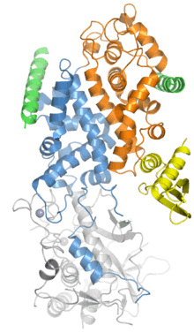
The Microprocessor complex consists minimally of two proteins: Drosha, a ribonuclease III enzyme; and DGCR8, a double-stranded RNA binding protein.[3][4][5] (DGCR8 is the name used in mammalian genetics, abbreviated from "DiGeorge syndrome critical region 8"; the homologous protein in model organisms such as flies and worms is called Pasha, for Partner of Drosha.) The stoichiometry of the minimal complex has been experimentally difficult to determine, but has been determined by biochemical analysis, single-molecule experiments, and X-ray crystallography to be a heterotrimer of two DGCR8 proteins to one Drosha.[6][7][8]
In addition to the minimal catalytically active Microprocessor components, additional cofactors such as DEAD box RNA helicases and heterogeneous nuclear ribonucleoproteins may be present in the complex to mediate the activity of Drosha.[3] Some miRNAs are processed by Microprocessor only in the presence of specific cofactors.[9]
Function
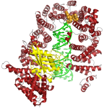
Located in the cell nucleus, the complex cleaves primary miRNA (pri-miRNA), typically at least 1000 nucleotides long, into precursor miRNA (pre-miRNA) molecules of around 70 nucleotides containing a stem-loop or hairpin structure. Pri-miRNA substrates can be derived either from non-coding RNA genes or from introns. In the latter case, there is evidence that the Microprocessor complex interacts with the spliceosome and that the pri-miRNA processing occurs prior to splicing.[4][10]
DGCR8 recognizes the junctions between hairpin structures and single-stranded RNA and serves to orient Drosha to cleave around 11 nucleotides away from the junctions. Microprocessor cleavage of pri-miRNAs typically occurs co-transcriptionally[11] and leaves a characteristic RNase III single-stranded overhang of 2-3 nucleotides, which serves as a recognition element for the transport protein exportin-5. Pre-miRNAs are exported from the nucleus to the cytoplasm in a RanGTP-dependent manner and are further processed, typically by the endoribonuclease enzyme Dicer.[3][4][5]
Although the large majority of miRNAs undergo processing by Microprocessor, a small number of exceptions called mirtrons have been described; these are very small introns which, after splicing, have the appropriate size and stem-loop structure to serve as a pre-miRNA.[12] The processing pathways for microRNA and for exogenously derived small interfering RNA converge at the point of Dicer processing and are largely identical downstream. Broadly defined, both pathways constitute RNA interference.[4][12]
Regulation
Gene regulation by miRNA is widespread across many genomes - by some estimates more than 60% of human protein-coding genes are likely to be regulated by miRNA,[13] though the quality of experimental evidence for miRNA-target interactions is often weak.[14] Because processing by Microprocessor is a major determinant of miRNA abundance, Microprocessor itself is then an important target of regulation. Both Drosha and DGCR8 are subject to regulation by post-translational modifications modulating stability, intracellular localization, and activity levels. Activity against particular substrates may be regulated by additional protein cofactors interacting with the Microprocessor complex. The loop region of the pri-miRNA stem-loop is also a recognition element for regulatory proteins, which may up- or down-regulate Microprocessor processing of the specific miRNAs they target.[9]
Microprocessor itself is autoregulated by negative feedback through association with a pri-miRNA-like hairpin structure found in the DGCR8 mRNA, which when cleaved reduces DGCR8 expression. The structure in this case is located in an exon and is unlikely to itself function as miRNA in its own right.[9]
Evolution
Drosha shares striking structural similarity with the downstream ribonuclease Dicer, suggesting an evolutionary relationship, through Drosha and related enzymes are found only in animals while Dicer relatives are widely distributed, including among protozoans.[8] Both components of Microprocessor are conserved among the vast majority of metazoans with known genomes. Mnemiopsis leidyi, a ctenophore, lacks both Drosha and DGCR8 homologs, as well as recognizable miRNAs, and is the only known metazoan with no detectable genomic evidence of Drosha.[15] In plants, the miRNA biogenesis pathway is somewhat different; neither Drosha nor DGCR8 has a homolog in plant cells, where the first step in miRNA processing is usually executed by a different nuclear ribonuclease, DCL1, a homolog of Dicer.[9][16]
It has been suggested based on phylogenetic analysis that the key components of RNA interference based on exogenous substrates were present in the ancestral eukaryote, likely as an immune mechanism against viruses and transposable elements. Elaboration of this pathway for miRNA-mediated gene regulation is thought to have evolved later.[17]
References
- ↑ Gregory, RI; Yan, KP; Amuthan, G; Chendrimada, T; Doratotaj, B; Cooch, N; Shiekhattar, R (11 November 2004). "The Microprocessor complex mediates the genesis of microRNAs.". Nature. 432 (7014): 235–40. doi:10.1038/nature03120. PMID 15531877.
- ↑ Denli, AM; Tops, BB; Plasterk, RH; Ketting, RF; Hannon, GJ (11 November 2004). "Processing of primary microRNAs by the Microprocessor complex.". Nature. 432 (7014): 231–5. doi:10.1038/nature03049. PMID 15531879.
- 1 2 3 4 Siomi, H; Siomi, MC (14 May 2010). "Posttranscriptional regulation of microRNA biogenesis in animals.". Molecular Cell. 38 (3): 323–32. doi:10.1016/j.molcel.2010.03.013. PMID 20471939.
- 1 2 3 4 5 Wilson, RC; Doudna, JA (2013). "Molecular mechanisms of RNA interference.". Annual Review of Biophysics. 42: 217–39. doi:10.1146/annurev-biophys-083012-130404. PMID 23654304.
- 1 2 3 Macias, S; Cordiner, RA; Cáceres, JF (August 2013). "Cellular functions of the microprocessor.". Biochemical Society Transactions. 41 (4): 838–43. doi:10.1042/BST20130011. PMID 23863141.
- ↑ Herbert, KM; Sarkar, SK; Mills, M; Delgado De la Herran, HC; Neuman, KC; Steitz, JA (February 2016). "A heterotrimer model of the complete Microprocessor complex revealed by single-molecule subunit counting.". RNA (New York, N.Y.). 22 (2): 175–83. doi:10.1261/rna.054684.115. PMID 26683315.
- ↑ Nguyen, TA; Jo, MH; Choi, YG; Park, J; Kwon, SC; Hohng, S; Kim, VN; Woo, JS (4 June 2015). "Functional Anatomy of the Human Microprocessor.". Cell. 161 (6): 1374–87. doi:10.1016/j.cell.2015.05.010. PMID 26027739.
- 1 2 Kwon, SC; Nguyen, TA; Choi, YG; Jo, MH; Hohng, S; Kim, VN; Woo, JS (14 January 2016). "Structure of Human DROSHA.". Cell. 164 (1-2): 81–90. doi:10.1016/j.cell.2015.12.019. PMID 26748718.
- 1 2 3 4 Ha, M; Kim, VN (August 2014). "Regulation of microRNA biogenesis.". Nature reviews. Molecular cell biology. 15 (8): 509–24. doi:10.1038/nrm3838. PMID 25027649.
- ↑ Kataoka, N; Fujita, M; Ohno, M (June 2009). "Functional association of the Microprocessor complex with the spliceosome.". Molecular and Cellular Biology. 29 (12): 3243–54. doi:10.1128/MCB.00360-09. PMC 2698730
 . PMID 19349299.
 - ↑ Morlando, M; Ballarino, M; Gromak, N; Pagano, F; Bozzoni, I; Proudfoot, NJ (September 2008). "Primary microRNA transcripts are processed co-transcriptionally.". Nature Structural & Molecular Biology. 15 (9): 902–9. doi:10.1038/nsmb.1475. PMID 19172742.
- 1 2 Winter, J; Jung, S; Keller, S; Gregory, RI; Diederichs, S (March 2009). "Many roads to maturity: microRNA biogenesis pathways and their regulation.". Nature Cell Biology. 11 (3): 228–34. doi:10.1038/ncb0309-228. PMID 19255566.
- ↑ Friedman, RC; Farh, KK; Burge, CB; Bartel, DP (January 2009). "Most mammalian mRNAs are conserved targets of microRNAs.". Genome Research. 19 (1): 92–105. doi:10.1101/gr.082701.108. PMC 2612969. PMID 18955434.
- ↑ Lee, YJ; Kim, V; Muth, DC; Witwer, KW (November 2015). "Validated MicroRNA Target Databases: An Evaluation.". Drug development research. 76 (7): 389–96. doi:10.1002/ddr.21278. PMID 26286669.
- ↑ Maxwell, EK; Ryan, JF; Schnitzler, CE; Browne, WE; Baxevanis, AD (20 December 2012). "MicroRNAs and essential components of the microRNA processing machinery are not encoded in the genome of the ctenophore Mnemiopsis leidyi.". BMC Genomics. 13: 714. doi:10.1186/1471-2164-13-714. PMC 3563456. PMID 23256903.
- ↑ Axtell, MJ; Westholm, JO; Lai, EC (2011). "Vive la différence: biogenesis and evolution of microRNAs in plants and animals.". Genome Biology. 12 (4): 221. doi:10.1186/gb-2011-12-4-221. PMC 3218855. PMID 21554756.
- ↑ Cerutti, H; Casas-Mollano, JA (August 2006). "On the origin and functions of RNA-mediated silencing: from protists to man.". Current genetics. 50 (2): 81–99. doi:10.1007/s00294-006-0078-x. PMC 2583075. PMID 16691418.