Bone healing

Bone healing, or fracture healing, is a proliferative physiological process in which the body facilitates the repair of a bone fracture.
Generally bone fracture treatment consists of a doctor reducing (pushing) displaced bones back into place via relocation with or without anaesthetic, stabilizing their position to aid union, and then waiting for the bone's natural healing process to occur.
Adequate nutrient intake has been found to significantly affect the integrity of the fracture repair.[1] Age, Bone type, drug therapy and pre existing bone pathology are factors which affect healing. The role of bone healing is to produce new bone without a scar as seen in other tissues which would be a structural weakness or deformity.[2]
The process of the entire regeneration of the bone can depend on the angle of dislocation or fracture. While the bone formation usually spans the entire duration of the healing process, in some instances, bone marrow within the fracture has healed two or fewer weeks before the final remodeling phase.
While immobilization and surgery may facilitate healing, a fracture ultimately heals through physiological processes. The healing process is mainly determined by the periosteum (the connective tissue membrane covering the bone). The periosteum is one source of precursor cells which develop into chondroblasts and osteoblasts that are essential to the healing of bone. The bone marrow (when present), endosteum, small blood vessels, and fibroblasts are other sources of precursor cells.[3]
Phases
There are three major phases of fracture healing,[4] two of which can be further sub-divided to make a total of five phases:
- 1. Reactive phase
- i. Fracture and inflammatory phase
- ii. Granulation tissue formation
- 2. Reparative phase
- iii. Cartilage callus formation
- iv. Lamellar bone deposition
- 3. Remodeling phase
- v. Remodeling to original bone contour
Reactive
After fracture, the first change seen by light and electron microscopy is the presence of blood cells within the tissues adjacent to the injury site. Soon after fracture, the blood vessels constrict, stopping any further bleeding.[5] Within a few hours after fracture, the extravascular blood cells form a blood clot, known as a hematoma. These cells release cytokines and increase blood capillary permeability. All of the cells within the blood clot degenerate and die.[6] Some of the cells outside of the blood clot, but adjacent to the injury site, also degenerate and die.[7] Within this same area, the fibroblasts survive and replicate. They form a loose aggregate of cells, interspersed with small blood vessels, known as granulation tissue.[8] This tissue reduces strain across the fracture site. Osteoclasts move in to reabsorb dead bone ends and other necrotic tissue are removed.[9]
Reparative
Days after fracture, the cells of the periosteum replicate and transform. The periosteal cells proximal (closest) to the fracture gap develop into chondroblasts which form hyaline cartilage. The periosteal cells distal to (further from) the fracture gap develop into osteoblasts which form woven bone. The fibroblasts within the granulation tissue develop into chondroblasts which also form hyaline cartilage.[10] These two new tissues grow in size until they unite with their counterparts from other parts of the fracture. These processes culminate in a new mass of heterogeneous tissue which is known as the fracture callus.[11] Eventually, the fracture gap is bridged by the hyaline cartilage and woven bone, restoring some of its original strength.
The next phase is the replacement of the hyaline cartilage and woven bone with lamellar bone. The replacement process is known as endochondral ossification with respect to the hyaline cartilage and bony substitution with respect to the woven bone. Substitution of the woven bone with lamellar bone precedes the substitution of the hyaline cartilage with lamellar bone. The lamellar bone begins forming soon after the collagen matrix of either tissue becomes mineralized. At this point, the mineralized matrix is penetrated by channels, each containing a microvessel and numerous osteoblasts. The osteoblasts form new lamellar bone upon the recently exposed surface of the mineralized matrix. This new lamellar bone is in the form of trabecular bone.[12] Eventually, all of the woven bone and cartilage of the original fracture callus is replaced by trabecular bone, restoring most of the bone's original strength.
Remodelling
The remodeling process substitutes the trabecular bone with compact bone. The trabecular bone is first resorbed by osteoclasts, creating a shallow resorption pit known as a "Howship's lacuna". Then osteoblasts deposit compact bone within the resorption pit. Eventually, the fracture callus is remodelled into a new shape which closely duplicates the bone's original shape and strength. The remodeling phase takes 3 to 5 years depending on factors such as age or general condition.[8] This process can be enhanced by certain synthetic injectable biomaterials, such as cerament, which are osteoconductive and actively promote bone healing.[13]
Obstructions to Bone Healing
_(14762515012).jpg)
- Poor blood supply which leads to the death of the osteocytes. Bone cell death is also dependent on degree of fracture and disruption to the Haversian system.
- Condition of the soft tissues. Soft tissue in between bone ends restrict healing.
- Nutrition and drug therapy. Poor general health reduces healing rate. Drugs that impair the inflammatory response impede healing also.
- Infection. Diverts the inflammatory response away from healing towards fighting of the infection.
- Age. Young bone unites more rapidly than adult bone.
- Pre existing Bone malignancy.
- Fracture healing is determined by mechanical factors and obstructions to healing include the bone not aligned and too much or little movement. Excess mobility can disrupt the bridging callus interfering with union. Slight biomechanical motion is also seen to improve callus formation.[9]
Complications
Complications of fracture healing include:
- Infection: this is the most common complication of fractures and predominantly occurs in open fractures. Post-traumatic wound infection is the most common cause of chronic osteomyelitis in patients. Osteomyelitis can also occur following surgical fixation of a fracture.[14]
- Non-union: no progression of healing within six months of a fracture occurring. The fracture pieces remain separated and can be caused by infection and/or lack of blood supply (Ischaemia) to the bone.[15] There are two types of non union atrophic and hypertrophic. Hypertrophic involves the formation of excess callus leading to bone ends appearing sclerotic causing a radiological "Elephants Foot" appearance. Atrophic non-union results in re-absorption and rounding of bone ends.[9]
- Mal-union: healing occurs but the healed bone has 'angular deformity, translation, or rotational alignment that requires surgical correction'. This is most common in long bones such as the femur.[16]
- Delayed union: healing times vary depending on the location of a fracture and the age of a patient. Delayed union is characterised by 'persistence of the fracture line and a scarcity or absence of callus formation' on x-ray. Healing is still occurring but at a much slower rate than normal.[15]
Gallery
-

Collagen fibers of woven bone
-

Osteoclast displaying many nuclei within its "foamy" cytoplasm
-
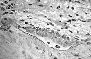
Osteoblasts forming compact bone, containing two osteocytes, within a resorption pit in trabecular bone
See also
Footnotes
- ↑ Susan E. Brown, PhD. "How to Speed Fracture Healing" (PDF). Center for Better Bones.
While no scientist has yet conducted a clinical trial using all 20 key nutrients for fracture healing, several studies have found multi-nutrient therapy to reduce complication and accelerate fracture healing.
- ↑ Gomez-Barrena E, Rosset P, Lozano D, Stanovici J, Ermthaller C, Gerbhard F. Bone fracture healing: Cell therapy in delayed unions and nonunions. Bone. 2015;70:93–101.
- ↑ Ferretti C, Mattioli-Belmonte M. Periosteum derived stem cells for regenerative medicine proposals: Boosting current knowledge. World Journal of Stem Cells. 2014;6(3):266-277. doi:10.4252/wjsc.v6.i3.266.
- ↑ Iain H. Kalfas, MD (2001). "Principles of Bone Healing". WebMD LLC.
- ↑ Brighton and Hunt (1997), p. 248: The extravascular blood cells are identified as erythrocytes, platelets and neutrophils.
- ↑ Brighton and Hunt (1991), p. 837: The cells within the clot are identified.
- ↑ Brighton and Hunt (1997)
- 1 2 Ham and Harris
- 1 2 3 Nyary Tamas, Scamell BE. (2015). Principles of bone and joint injuries and their healing. Surgery(Oxford). 33 (1), p 7-14.
- ↑ Brighton and Hunt (1997), p. 248: Two light micrographs showing the cells of the woven bone and hyaline cartilage.
- ↑ Brighton and Hunt (1986), p. 704: Two light micrographs of a typical fracture callus: one showing the tissues and the other showing the cells.
- ↑ Brighton and Hunt (1986); Brighton and Hunt (1997); Ham and Harris
- ↑ Hatten Jr., H.P. and Voor, J. (2012): Bone Healing Using a Bi-Phasic Ceramic Bone Substitute Demonstrated in Human Vertebroplasty and with Histology in a Rabbit Cancellous Bone Defect Model. Interventional Neuroradiology, vol. 18, pp. 105-113.
- ↑ Rowbotham, Emma; Barron, Dominic (2009). "Radiology of fracture complications". Orthopaedics and Trauma. 23 (1): 52–60. doi:10.1016/j.mporth.2008.12.008.
- 1 2 Jahagirdar, Rajeev; Scammell, Brigitte E (2008). "Principles of fracture healing and disorders of bone union". Surgery. 27 (2): 63–69. doi:10.1016/j.mpsur.2008.12.011.
- ↑ Chen, Andrew T; Vallier, Heather A (2016). "Noncontiguous and open fractures of the lower extremity: Epidemiology, complications, and unplanned procedures". Injury. 47 (3): 742–747. doi:10.1016/j.injury.2015.12.013.
References
- Brighton, Carl T. and Robert M. Hunt (1986), "Histochemical localization of calcium in the fracture callus with potassium pyroantimonate: possible role of chondrocyte mitochondrial calcium in callus calcification", Journal of Bone and Joint Surgery, 68-A (5): 703-715
- Brighton, Carl T. and Robert M. Hunt (1991), "Early histologic and ultrastructural changes in medullary fracture callus", Journal of Bone and Joint Surgery, 73-A (6): 832-847
- Brighton, Carl T. and Robert M. Hunt (1997), "Early histologic and ultrastructural changes in microvessels of periosteal callus", Journal of Orthopaedic Trauma, 11 (4): 244-253
- Ham, Arthur W. and William R. Harris (1972), "Repair and transplantation of bone", The biochemistry and physiology of bone, New York: Academic Press, p. 337-399
