Tibia
| Tibia | |
|---|---|
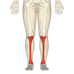 Position of tibia (shown in red) | |
 Cross section of the leg showing the different compartments (latin terminology) | |
| Details | |
| Articulations |
Knee, ankle, superior and inferior tibiofibular joint |
| Identifiers | |
| Latin | (os) tibia |
| MeSH | A02.835.232.043.650.883 |
| TA | A02.5.06.001 |
| FMA | 24476 |
The tibia /ˈtɪbiə/ (plural tibiae /ˈtɪbii/ or tibias), also known as the shinbone or shankbone, is the larger and stronger of the two bones in the leg below the knee in vertebrates (the other being the fibula), and it connects the knee with the ankle bones. The tibia is found on the medial side of the leg next to the fibula, and closer to the median plane or centre-line. The tibia is connected to the fibula by the interosseous membrane of the leg, forming a type of fibrous joint called a syndesmosis with very little movement. The tibia is named for the flute tibia. It is the second largest bone in the human body next to the femur. The leg bones are the strongest long bones as they support the rest of the body.
Structure
In human anatomy the tibia is the second largest bone next to the femur. As in other vertebrates the tibia is one of two bones in the lower leg, the other being the fibula, and is a component of the knee and ankle joints. The leg bones (femur, tibia and fibula) are the strongest long bones as they have to support the rest of the body.
The ossification or formation of the bone starts from three centers; one in the shaft and one in each extremity.
The tibia is categorized as a long bone and is as such composed of a diaphysis and two epiphyses. The diaphysis is the midsection of the tibia also known as shaft or body. While the epiphyses are the two rounded extremities of the bone; an upper (also known as superior or proximal) closest to the thigh and a lower (also known as inferior or distal) closest to the foot. The tibia is most contracted in the lower third and the distal extremity is smaller than the proximal.
Features
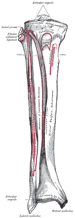 Muscle attachments (seen from front) | 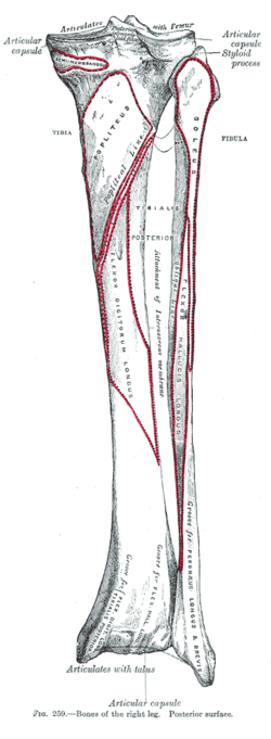 Muscle attachments (seen from back) |
The proximal or upper extremity of the tibia is expanded in the transverse plane with a medial and lateral condyle, which are both flattened in the horizontal plane. The medial condyle is the largest of the two and is better supported over the shaft. The upper surfaces of the condyles articulates with the femur to form the tibiofemoral joint, the weightbearing part of the kneejoint.[1]
The medial and lateral condyle are separated by the intercondylar area, where the cruciate ligaments and the menisci attach. Here the medial and lateral intercondylar tubercle forms the intercondylar eminence. Together with the medial and lateral condyle the intercondylar region forms the tibial plateau, which both articulates with and is anchored to the lower extremity of the femur. The intercondylar eminence divides the intercondylar area into an anterior and posterior part. The anterolateral region of the anterior intercondylar area are perforated by numerous small openings for nutrient arteries.[1] The articular surfaces of both condyles are concave, particularly centrally. The flatter outer margins are in contact with the menisci. The medial condyles superior surface is oval in form and extends laterally onto the side of medial intercondylar tubercle. The lateral condyles superior surface is more circular in form and its medial edge extends onto the side of the lateral intercondylar tubercle. The posterior surface of the medial condyle bears a horizontal groove for part of the attachment of the semimembranosus muscle, whereas the lateral condyle has a circular facet for articulation with the head of fibula.[1] Beneath the condyles is the tibial tuberosity which serves for attachment of the patellar ligament, a continuation of the quadriceps femoris muscle.[1]
The shaft or body of the tibia is triangular in cross-section and forms three borders: An anterior, medial and lateral or interosseous border. These three borders form three surfaces; the medial, lateral and posterior.[2] The forward flat part of the tibia is called the fibia, often confused with the fibula.
The lower extremity of the tibia is much smaller than the upper extremity and presents five surfaces; it is prolonged downward on its medial side as a strong pyramidal process, the medial malleolus. The lower extremity of the tibia together with the fibula and talus forms the ankle joint.
Blood supply
The tibia is supplied with blood from two sources: A nutrient artery, as the main source, and periosteal vessels derived from the anterior tibial artery.[3]
Joints
The tibia is a part of four joints; the knee, ankle, superior and inferior tibiofibular joint.
In the knee the tibia forms one of the two articulations with the femur, often referred to as the tibiofemoral components of the knee joint.[4][5] This is the weightbearing part of the knee joint.[2] The tibiofibular joints are the articulations between the tibia and fibula which allows very little movement. The proximal tibiofibular joint is a small plane joint. The joint is formed between the undersurface of the lateral tibial condyle and the head of fibula. The joint capsule is reinforced by anterior and posterior ligament of the head of the fibula.[2] The distal tibiofibular joint (tibiofibular syndesmosis) is formed by the rough, convex surface of the medial side of the distal end of the fibula, and a rough concave surface on the lateral side of the tibia.[2] The part of the ankle joint known as the talocrural joint, is a synovial hinge joint that connects the distal ends of the tibia and fibula in the lower limb with the proximal end of the talus. The articulation between the tibia and the talus bears more weight than between the smaller fibula and the talus.
Development


The tibia is ossified from three centers; a primary center for the diaphysis (shaft) and a secondary center for each epiphysis (extremity). Ossification begins in the center of the body, about the seventh week of fetal life, and gradually extends toward the extremities.
The center for the upper epiphysis appears before or shortly after birth at close to 34 weeks gestation; it is flattened in form, and has a thin tongue-shaped process in front, which forms the tuberosity; that for the lower epiphysis appears in the second year.
The lower epiphysis fuses with the tibial shaft at about the eighteenth, and the upper one fuses about the twentieth year.
Two additional centers occasionally exist, one for the tongue-shaped process of the upper epiphysis, which forms the tuberosity, and one for the medial malleolus.
Function
Muscle attachments
| Muscle | Direction | Attachment[6] |
| Tensor fasciae latae muscle | Insertion | Gerdy's tubercle |
| Quadriceps femoris muscle | Insertion | Tuberosity of the tibia |
| Sartorius muscle | Insertion | Pes anserinus |
| Gracilis muscle | Insertion | Pes anserinus |
| Semitendinosus muscle | Insertion | Pes anserinus |
| Horizontal head of the semimembranosus muscle | Insertion | Medial condyle |
| Popliteus muscle | Insertion | Posterior side of the tibia over the soleal line |
| Tibialis anterior muscle | Origin | Lateral side of the tibia |
| Extensor digitorum longus muscle | Origin | Lateral condyle |
| Soleus muscle | Origin | Posterior side of the tibia under the soleal line |
| Flexor digitorum longus muscle | Origin | Posterior side of the tibia under the soleal line |
Strength
The tibia has been modeled as taking an axial force during walking that is up to 4.7 bodyweight. Its bending moment in the sagittal plane in the late stance phase is up to 71.6 bodyweight times millimetre.[7]
Clinical significance
Fracture
Fractures of the tibia can be divided into those that only involve the tibia; bumper fracture, Segond fracture, Gosselin fracture, Toddler's fracture, and those including both the tibia and fibula; trimalleolar fracture, bimalleolar fracture, Pott's fracture.
Society and culture
In Judaism, the tibia, or shankbone, of a goat is used in the Passover Seder plate.
Other animals
The structure of the tibia in most other tetrapods is essentially similar to that in humans. The tuberosity of the tibia, a crest to which the patellar ligament attaches in mammals, is instead the point for the tendon of the quadriceps muscle in reptiles, birds, and amphibians, which have no patella.[8]
Additional images
 Shape of right tibia
 Longitudinal section of tibia showing interior
 Right knee joint from the front, showing interior ligaments
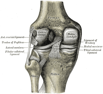 Left knee joint from behind, showing interior ligaments
 Left talocrural joint
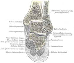 Coronal section through right talocrural and talocalcaneal joints
See also
References
This article incorporates text in the public domain from the 20th edition of Gray's Anatomy (1918)
- 1 2 3 4 Drake, Richard L.; Vogl, A. Wayne; Mitchell, Adam W. M. (2010). Gray´s Anatomy for Students (2nd ed.). pp. 558–560. ISBN 978-0-443-06952-9.
- 1 2 3 4 Drake, Richard L.; Vogl, A. Wayne; Mitchell, Adam W. M. (2010). Gray´s Anatomy for Students (2nd ed.). pp. 584–588. ISBN 978-0-443-06952-9.
- ↑ Nelson G, Kelly P, Peterson L, Janes J (1960). "Blood supply of the human tibia". J Bone Joint Surg Am. 42–A: 625–36. PMID 13854090.
- ↑ Rytter S, Egund N, Jensen LK, Bonde JP (2009). "Occupational kneeling and radiographic tibiofemoral and patellofemoral osteoarthritis". J Occup Med Toxicol. 4 (1): 19. doi:10.1186/1745-6673-4-19. PMC 2726153
 . PMID 19594940.
 - ↑ Gill TJ, Van de Velde SK, Wing DW, Oh LS, Hosseini A, Li G (December 2009). "Tibiofemoral and patellofemoral kinematics after reconstruction of an isolated posterior cruciate ligament injury: in vivo analysis during lunge". Am J Sports Med. 37 (12): 2377–85. doi:10.1177/0363546509341829. PMC 3832057. PMID 19726621.
- ↑ Bojsen-Møller, Finn; Simonsen, Erik B.; Tranum-Jensen, Jørgen (2001). Bevægeapparatets anatomi [Anatomy of the Locomotive Apparatus] (in Danish) (12th ed.). pp. 364–367. ISBN 978-87-628-0307-7.
- ↑ Wehner, T; Claes, L; Simon, U (2009). "Internal loads in the human tibia during gait". Clin Biomech. 24 (3): 299–302. doi:10.1016/j.clinbiomech.2008.12.007. PMID 19185959.
- ↑ Romer, Alfred Sherwood; Parsons, Thomas S. (1977). The Vertebrate Body. Philadelphia, PA: Holt-Saunders International. p. 205. ISBN 0-03-910284-X.
External links
| Wikimedia Commons has media related to Tibia. |
| Look up Tibia or shinbone in Wiktionary, the free dictionary. |