Spheroplast
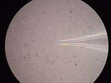
A spheroplast is a cell from which the cell wall has been almost completely removed, as by the action of penicillin. The name stems from the fact that after a microbe's cell wall is digested, membrane tension causes the cell to acquire a characteristic spherical shape. Spheroplasts are osmotically fragile, and will lyse if transferred to a hypotonic solution.
Uses and applications
Antibiotic discovery
From the 1960s into the 1990s Merck and Co. used a spheroplast screen as a primary method for discovery of antibiotics that inhibit cell wall biosynthesis. In this screen devised by Eugene Dulaney, growing bacteria were exposed to test substances under hypertonic conditions. Inhibitors of cell wall synthesis caused growing bacteria to form spheroplasts. This screen enabled the discovery of fosfomycin, cephamycin C, thienamycin and several carbapenems.[1]
Patch clamping
Specially prepared giant spheroplasts of Gram-negative bacteria can be used to study the function of bacterial ion channels through a technique called patch clamp, which was originally designed for characterizing the behavior of neurons and other excitable cells. To prepare giant spheroplasts, bacteria are grown in a medium containing chemicals that prevent the cells from dividing completely. This causes bacteria to form long "snakes" that share a single membrane and cytoplasm. After a period of time, the cell walls of the "snakes" are digested, and the bacteria collapse into very large spheres surrounded by a single lipid bilayer. The membrane can then be analyzed on a patch clamp apparatus to determine the phenotype of the ion channels embedded in it. It is also common to overexpress a particular channel to amplify its effect and make it easier to characterize.
The technique of patch clamping giant E. coli spheroplasts has been used extensively for studying the native mechanosensitive channels (MscL, MscS, and MscM) of E. coli since 1987.[2][3] Recently, it has been extended to study other heterologously expressed ion channels and it has been shown that the giant E. coli spheroplast can be used as an ion-channel expression system comparable to the Xenopus oocyte.[4][5][6][7]
Cell lysis
Yeast cells are normally protected by a thick cell wall which makes extraction of cellular proteins difficult. Enzymatic digestion of the cell wall with zymolyase, creating spheroplasts, renders the cells vulnerable to easy lysis with detergents or rapid osmolar pressure changes.
Transfection
Bacterial spheroplasts, with suitable recombinant DNA inserted into it, can be used to transfect animal cells. Spheroplasts with recombinant DNA are introduced into the media containing animal cells and are fused by polyethylene glycol (PEG). With this methodology, nearly 100% of the animal cells may take up the foreign DNA.
See also
References
- ↑ Silver, L.L. Rational approaches to antibiotic discovery: pre-genomic directed and phenotypic screening, 2.4.2 Screens for spheroplast formation. In: Thomas Dougherty, Michael J. Pucci, Antibiotic Discovery and Development. Chap. 2, p. 46.
- ↑ Martinac, B., Buechner, M., Delcour, A. H., Adler, J., and Kung, C. (1987) Pressure-sensitive ion channel in Escherichia coli. Proc. Natl. Acad. Sci. USA 84, 2297-2301.
- ↑ Blount, P., Sukharev, S. I., Moe, P. C., Martinac, B., and Kung, C. (1999) Mechanosensitive channels of bacteria. Methods in Enzymology 294, 458-482.
- ↑ Santos, J. S., Lundby, A., Zazueta, C., and Montal, M. (2006) Molecular template for a voltage sensor in a novel K+ channel. I. Identification and functional characterization of KvLm, a voltage-gated K+ channel from Listeria monocytogenes. Journal of General Physiology 128(3), 283-292.
- ↑ Nakayama, Y., Fujiu, K., Sokabe, M., and Yoshimura, K. (2007) Molecular and electrophysiological characterization of a mechanosensitive channel expressed in the chloroplasts of Chlamydomonas. Proc. Natl. Acad. Sci. USA 104, 5883-5888.
- ↑ Kuo, M. M.-C., Baker, K. A., Wong, L., and Choe, S. (2007) Dynamic oligomeric conversions of the cytoplasmic RCK domains mediate MthK potassium channel activity. Proc. Natl. Acad. Sci. USA 104, 2151-2156.
- ↑ Kuo, M. M.-C., Saimi, Y., Kung, C., and Choe, S. (2007). Patch-clamp and phenotypic analyses of a prokaryotic cyclic nucleotide-gated K+ channel using Escherichia Coli as a host. J. Biol. Chem. 282, 24294-24301.
External links
- Spheroplasts at the US National Library of Medicine Medical Subject Headings (MeSH)