Pulp capping


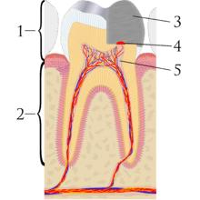
Pulp capping is a technique used in dental restorations to prevent the dental pulp from dying, after being exposed, or nearly exposed during a cavity preparation. When dental caries is removed from a tooth, all or most of the infected and softened enamel and dentin are removed. This can lead to the pulp of the tooth either being exposed or nearly exposed which causes pulpitis (inflammation). Pulpitis, in turn, can become irreversible, leading to pain and pulp necrosis, and necessitating either root canal treatment or extraction.[1] The ultimate goal of pulp capping or stepwise caries removal is to protect a healthy dental pulp and avoid the need for root canal therapy.
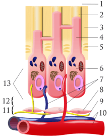
To prevent the pulp from deteriorating when a dental restoration gets near the pulp, the dentist will place a small amount of a sedative dressing, such as calcium hydroxide or MTA. These materials, protect the pulp from noxious agents (heat, cold, bacteria) and stimulate the cell-rich zone of the pulp to lay down a bridge of reparative dentin. Dentin formation usually starts within 30 days of the pulp capping (there can be a delay in onset of dentin formation if the odontoblasts of the pulp are injured during cavity removal) and is largely completed by 130 days.[2]:491–494
2 different types of pulp cap are distinguished. In direct pulp capping, the protective dressing is placed directly over an exposed pulp; and in indirect pulp capping, a thin layer of softened dentin, that if removed would expose the pulp, is left in place and the protective dressing is placed on top.[3] A direct pulp cap is a one-stage procedure, whereas an indirect pulp cap is a 2-stage procedure over about 6 months.
Direct pulp cap
This technique is used when a pulpal exposure occurs, either due to caries extending to the pulp chamber, or accidentally, during caries removal. It is only feasible if the exposure is made through non infected dentin and there is no recent history of spontaneous pain (i.e. irreversible pulpitis) and a bacteria-tight seal can be applied.[3] Once the exposure is made, the tooth is isolated from saliva to prevent contamination by use of a dental dam, if it was not already in place. The tooth is then washed and dried, and the protective material placed, followed finally by a dental restoration which gives a bacteria-tight seal to prevent infection. Since pulp capping is not always successful in maintaining the vitality of the pulp, the dentist will usually keep the status of the tooth under review for about 1 year after the procedure.[3]
Indirect pulp cap (stepwise caries removal)
This technique is used when most of the decay has been removed from a deep cavity, but some softened dentin and decay remains over the pulp chamber that if removed would expose the pulp and trigger irreversible pulpitis. Instead, the dentist intentionally leaves the softened dentin/decay in place, and uses a layer of protective temporary material which promotes remineralization of the softened dentin over the pulp and the laying down of new layers of tertiary dentin in the pulp chamber. A temporary filling is used to keep the material in place, and about 6 months later, the cavity is re-opened and hopefully there is now enough sound dentin over the pulp (a "dentin bridge") that any residual softened dentin can be removed and a permanent filling can be placed. This method is also called "stepwise caries removal."[3][4]
See also
References
- ↑ Stockton, LW (1999). "Vital Pulp Capping: A Worthwhile Procedure (review)". J Can Dent Assoc. 65: 328–31.
- ↑ Hargreaves, K (2011). Cohen's Pathways of the Pulp, Tenth Edition. St. Louis, Missouri: Mosby Elsevier. ISBN 978-0-323-06489-7.
- 1 2 3 4 "Quality guidelines for endodontic treatment: consensus report of the European Society of Endodontology". International Endodontic Journal. 39 (12): 921–930. December 2006. doi:10.1111/j.1365-2591.2006.01180.x.
- ↑ Schwendicke F, Dörfer CE, Paris S (Apr 2013). "Incomplete caries removal: a systematic review and meta-analysis.". J Dent Res. 92 (4): 306–14. doi:10.1177/0022034513477425. PMID 23396521.