Posterolateral sulcus of medulla oblongata
| Posterolateral sulcus of medulla oblongata | |
|---|---|
 Hind- and mid-brains; postero-lateral view. | |
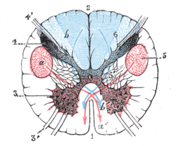 Section of the medulla oblongata through the lower part of the decussation of the pyramids.
the blue arrow, b, b’, indicates the course which the sensory fibers take. | |
| Details | |
| Identifiers | |
| Latin | sulcus posterolateralis medullae oblongatae |
| NeuroNames | hier-703 |
| TA | A14.1.04.012 |
| FMA | 75608 |
The accessory, vagus, and glossopharyngeal nerves correspond with the posterior nerve roots, and are attached to the bottom of a sulcus named the posterolateral sulcus (or dorsolateral sulcus).
Additional images
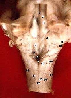 Human caudal brainstem posterior view description
References
This article incorporates text in the public domain from the 20th edition of Gray's Anatomy (1918)
This article is issued from Wikipedia - version of the 11/1/2016. The text is available under the Creative Commons Attribution/Share Alike but additional terms may apply for the media files.