Porcine reproductive and respiratory syndrome virus
| Arterivirus | |
|---|---|
| Virus classification | |
| Group: | Group IV ((+)ssRNA) |
| Order: | Nidovirales |
| Family: | Arteriviridae |
| Genus: | Arterivirus |
| Species: | Porcine reproductive and respiratory syndrome virus |
Porcine reproductive and respiratory syndrome virus (PRRSV) is a virus that causes a disease of pigs, called porcine reproductive and respiratory syndrome (PRRS), also known as blue-ear pig disease (in Chinese, zhū láněr bìng 豬藍耳病). This economically important, panzootic disease causes reproductive failure in breeding stock and respiratory tract illness in young pigs. Initially referred to as "mystery swine disease" and "mystery reproductive syndrome," it was first reported in 1987 in North America (2) and Central Europe (3). The disease costs the United States swine industry around $644 million annually, and recent estimates in Europe found that it costs almost 1.5b€ every year.
Classification
PRRSV is a small, enveloped RNA virus. It contains a single-stranded, positive-sense, RNA genome with a size of approximately 15 kilobases. The genome contains nine open reading frames (Meulenburg et al., 1992, Lee and Yoo, 2005).
PRRSV is a member of the genus Arterivirus, family Arteriviridae, order Nidovirales.[1] The three other members of the genus Arterivirus are: equine arteritis virus, simian hemorrhagic fever virus, and lactate dehydrogenase elevating virus (4-5).
Strains
The two prototype strains of PRRSV are the North American strain, VR-2332, and the European strain, the Lelystad virus (LV). The European and North American PRRSV strains cause similar clinical symptoms, but represent two distinct viral genotypes whose genomes diverge by approximately 40% (6), thus creating a veil of mystery about the origin of this virus. The genetic variation among the viruses isolated from different places (7-8) increases the difficulty of developing vaccines against it. Similarly, maintaining diagnostic PCR detection assays is difficult due to the high mutation rate of this virus, see Risk of Missed PRRS PCR Detection.
In the early 2000s a highly pathogenic strain of the North American genotype emerged in China. This strain, HP-PRRSV, is more virulent than all other strains, and causes great losses in Asian countries worldwide. Later a study showed that accelerated evolution of a group of strains in China.[2]
Clinical signs
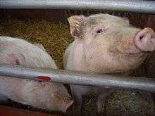
Subclinical infections are common, with clinical signs occurring sporadically in a herd. Clinical signs include reproductive failure in sows such as abortions and giving birth to stillborn or mummified fetuses, and cyanosis of the ear and vulva. In neonatal pigs, the disease causes respiratory distress, with increased susceptibility to respiratory infections such as Glasser's disease.
Laboratory diagnosis
Laboratory-based diagnostic tests have evolved significantly since initial discovery of the PRRS virus in the late 1980s. Initially viral culture was used to confirm PRRSV in serum or tissue samples. This process involves growing the virus in-vitro on cell lines over a period of 3–14 days or longer. If cytopathic effect is observed during culture, the culture is confirmed as the PRRS virus by direct fluorescent antibody or other confirmation method prior to reporting the sample as positive for presence of PRRSV.
In the late 1990s, nested PCR was used to the detect the virus as it showed improved sensitivity over non-nested PCR.[3] Now, quantitative PCR assays offered as-good or better sensitivity than nested PCR, fast turnaround time in the lab, and lower rates of cross-contamination via closed-tube amplification.
As an RNA virus with a 15 kb genome, PRRS mutates at a relatively high rate as it is transmitted from pig-pig over time.[4] The calculated rate of PRRSV nucleotide substitution is the highest reported so far for an RNA virus. It is estimated as 4.7-9.8 x 10⁻² / site / year.[5]
Though the quantitative PCR tests used now have high sensitivity and specificity, these improvements have come with some hazards as well. Quantitative PCR using Taq-man chemistry is prone to false-negative results when the virus mutates.[6][7][8] A false negative result occurs when a test fails to detect the presence of the virus. Studies have found that even a single base-pair change in the viral RNA under the labeled probe can cause failure of detection.[6] This specific source of the false-negative is not due to operator error on the part of the lab and is un-knowable at the time of testing.
The scenario that follows demonstrates how this hazard can result in risk to pork producers and laboratories:
→A strain of PRRS virus mutates during circulation within a herd. This strain spreads and becomes the predominant strain within the herd.
- →A veterinarian takes a random statistical sample of (let's say 30) animals within the herd, either in reaction to clinical signs or during routine health monitoring. Even though 30 animals are sampled, the mutant strain makes up the majority of PRRSV in all samples. The samples are submitted to the veterinary diagnostic lab for PRRS quantitative PCR testing in order to get a quick diagnosis.
- →Mutation in viral RNA occurred in the small region(s) of the virus that the probe binds to, so the lab finds no signal and reports samples as 'negative' for absence of PRRS virus.
- →The veterinarian found evidence of other etiologic agents from related samples sent to the lab and assumes these must be the cause of clinical signs on farm. The animal owners are told that they can resume shipments of piglets from the sampled farm to another site where 5,000 PRRS-naive animals reside.
- →As the virus circulates in the new herd, more copies of the mutant virus are circulated. Further sampling continues to result in PRRS-negative results, and eventually clinical signs cause the veterinarian to explore other PRRSV test methods. The lab is contacted when other methods confirm that PRRSV is causing the signs on-farm.
- →At this point, the lab may attempt to isolate the virus (1 week at best), sequence the RNA from it (1 week), and analyze the sequence for miss-matches with the TaqMan probes used in the detection assay (1 week). Now the assay probe must be re-designed to allow detection of this new variant while still remaining sensitive to all other known strains. Optimization and validation of the re-designed assay can then take a substantial amount of time.
- →Meanwhile, the index case herd can no longer utilize PCR to determine which management options to use to control virus spread. Until the test is updated and implemented, the veterinarian cannot continue to use the diagnostic lab for testing, so samples are sent elsewhere and confidence in the laboratory is diminished.
This series of events is a frustrating and expensive event for veterinarian, diagnostic lab, and animal owners. Many labs in the United States each use their own quantitative PCR method and communication of test failures due to new strains to other diagnostic labs is difficult. As a result, information learned about new strains is not leveraged across many diagnostic labs. Due to the cost of testing and rapid detection of new virus introduction, PCR alone is often relied on as the primary screening tool. This over-reliance on a single diagnostic assay (of which none are 100% sensitive and specific) lead to longer interval of virus spread while the problem is being resolved.
Veterinarian and producer
Veterinarians can reduce the impact of this risk by paying close attention to clinical signs and utilizing more than one PRRS diagnostic test. Early communication with the lab is essential as often other methods can quickly be employed on existing samples. Given the rate of mutation for the PRRS virus, contingency plans should be developed for false-negative events that include selection of alternative labs and tests.
Diagnostic laboratory
Some laboratories have moved to the use of commercially developed and maintained quantitative PCR assays, which transfers the work of assay updates to a 3rd party albeit at a significant extra cost over in-house developed assays. In recent years, this strategy has allowed quicker response to new variants than would have been previously possible (unpublished). By commercial manufacturers leveraging assay updates across multiple labs, it is possible that detection capabilities for all client labs is improved. The flip-side of this approach is that if all labs run the same assay, there are limited options for veterinarians when an alternate assay is quickly needed.
Earlier technologies such as nested PCR are often called on during an investigation if the lab has retained the capability to perform them. By using these earlier methods the laboratory staff are more quickly able to identify the new strain due to their more robust detection capabilities.
Control
Porcine Reproductive and Respiratory Syndrome (PRRS) is a complex disease. Modified Live Vaccines (MLV) vaccines are the primary immunological tool for its control, but PRRS control goes way beyond than just vaccination, and in order to achieve sustainable results, a systematic approach should be implemented. It requires a full understanding of the disease and a set of tools to achieve a long term success, therefore a standardized 5 step process has been developed to successfully achieve PRRS control. A strong platform to consolidate PRRS control in pig farms, large production systems and even geographical areas has been developed. This platform is a pig population approach having as main goals: - to maximize immunity, - reduce PRRS virus (PRRSv) exposure and - prevent new PRRSv infections. The Complexity of PRRS has allowed implementing successfully this methodology in the Swine Industry around the globe.
See also
References
- ↑ Balasuriya and Snijder (2008). "Arteriviruses". Animal Viruses: Molecular Biology. Caister Academic Press. ISBN 978-1-904455-22-6.
- ↑ Song J, Shen D, Cui J, Zhao B (Oct 2010). "Accelerated evolution of PRRSV during recent outbreaks in China". Virus Genes. 41 (2): 241–5. doi:10.1007/s11262-010-0507-2. PMID 20652733.
- ↑ Christopher-Hennings, Jane; et al. (July 1995). "Detection of Porcine Reproductive and Respiratory Syndrome Virus in Boar Semen by PCR". Journal of Clinical Microbiology. 33: 1730–1734. PMID 7665637.
- ↑ Shi, Mang; et al. (2010). "Molecular epidemiology of PRRSV: A phylogenic perspective.". Virus Research. 154: 7–17. doi:10.1016/j.virusres.2010.08.014.
- ↑ Murtaugh, Michael; et al. (2010). "The ever-expanding diversity of porcine reproductive and respiratory syndrome virus". Virus Research. 154: 18–30. doi:10.1016/j.virusres.2010.08.015.
- 1 2 Klungthong, Chonticha; et al. (2010). "The impact of primer and probe-template mismatches on the sensitivity of pandemic influenza A/H1N1/2009 virus detection by real-time RT-PCR". Journal of Clinical Virology. 48: 91–95. doi:10.1016/j.jcv.2010.03.012.
- ↑ Pyne, Michael; et al. (August 2010). "Evaluation of the Roche Cobas AmpliPrep/Cobas TaqMan HIV-1 Test and Identification of Rare Polymorphisms Potentially Affecting Assay Performance". Journal of Clinical Microbiology. 48: 2852–2858. doi:10.1128/JCM.00776-10. PMID 20573864.
- ↑ Toplak, I.; et al. (2011). "Identification of genetically diverse sequence of PRRSV in Slovenia and the impact on the sensitivity of four molecular tests". Journal of Virological Methods. 179: 51–56. doi:10.1016/j.jviromet.2011.09.019.
- Benfield D, Collins J, Dee S, Halbur P, Joo H, Lager K, et al. Porcine reproductive and respiratory syndrome. In: Straw BE, D’Allaire S, Mengeling WL, Taylor DJ, editors. Diseases of the swine. 8th ed. Ames, Iowa: Iowa State University Press; 1999. p. 201–32.
- Collins J, Benfield D, Christianson W, Harris L, Hennings J, Shaw D, et al. Isolation of swine infertility and respiratory syndrome virus (isolate ATCC VR-2332) in North America and experimental reproduction of the disease in gnotobiotic pigs. J Vet Diagn Invest 1992;4:117–26
- Wensvoort G. Lelystad virus and the porcine epidemic abortion and respiratory syndrome. Vet Res 1993;24:117–24
- Cavenagh D. Nidovirales: a new order comprising Coronaviridae and Arteriviridae. Arch Virol 1997;142:629–33
- Thiel HJ, Meyers G, Stark R, Tautz N, Rumenapf T, Unger G, Conzelmann KK., Molecular characterization of positive-strand RNA viruses: pestiviruses and the porcine reproductive and respiratory syndrome virus (PRRSV). Arch Virol Suppl. 1993;7:41-52
- Nelsen C, Murtaugh M, Faaberg K. Porcine reproductive and respiratory syndrome virus comparison: divergent evolution on two continents. J Virol 1999;73:270–80
- Kapur V, Elam MR, Pawlovich TM, Murtaugh MP. Genetic variation in porcine reproductive and respiratory syndrome virus isolates in the midwestern United States. J Gen Virol. 1996 Jun;77 ( Pt 6):1271-6
- Meng XJ, Paul PS, Halbur PG, Morozov I.Sequence comparison of open reading frames 2 to 5 of low and high virulence United States isolates of porcine reproductive and respiratory syndrome virus. J Gen Virol. 1995 Dec;76 ( Pt 12):3181-8
- Barboza, David. Chinese Pig Virus Causes Concern Around the Globe. The New York Times. August 15, 2007.
- Meulenberg, J. J.; Hulst, M. M.; de Meijer, E. J.; Moonen, P. L.; den Besten, A.; de Kluyver, E. P.; Wensvoort, G., and Moormann, R. J. Lelystad virus, the causative agent of porcine epidemic abortion and respiratory syndrome (PEARS), is related to LDV and EAV. Virology. 1993 Jan; 192(1):62-72.
- Lee, C. and Yoo, D. Cysteine residues of the porcine reproductive and respiratory syndrome virus small envelope protein are non-essential for virus infectivity. J Gen Virol. 2005 Nov; 86(11):3091-6.
External links
- www.prrs.com, The complete guide to PRRS understanding and control.
- OIE Q&A about the Porcine reproductive and respiratory syndrome
- New York Times article on the 2007 epidemic
- "Europe's piglets die in mystery plague". New Scientist. May 4, 1991. Retrieved 2007-06-28.
- Animal viruses
- PRRS Research Award for PRRS Eradication
- PADRAP Production Animal Disease Risk Assessment Program PRRS Risk Survey
- The latest new features on this swine disease, created by Scott A. Dee
- PRRS, from ThePigSite disease guide