Phonocardiogram
| Phonocardiogram | |
|---|---|
| Diagnostics | |
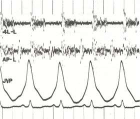 Phonocardiogram and jugular venous pulse tracing from a middle-aged man with pulmonary hypertension (pulmonary artery pressure 70 mm Hg) caused by cardiomyopathy. The jugular venous pulse tracing demonstrates a prominent a wave without a c or v wave being observed. The phonocardiograms (fourth left interspace and cardiac apex) show a murmur of tricuspid insufficiency and ventricular and atrial gallops.[1] | |
| ICD-9-CM | 89.55 |
A phonocardiogram (or PCG) is a plot of high-fidelity recording of the sounds and murmurs made by the heart with the help of the machine called the phonocardiograph; thus, phonocardiography is the recording of all the sounds made by the heart during a cardiac cycle. .[2]
Medical use
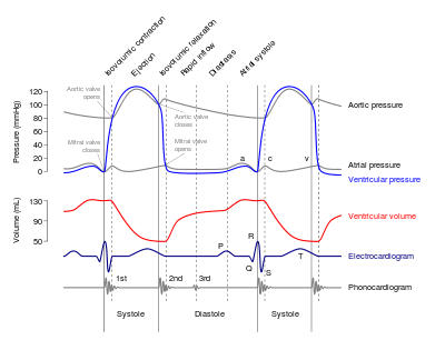
The sounds result from vibrations created by closure of the heart valves, there are at least two: the first when the atrioventricular valves close at the beginning of systole and the second when the aortic valve and pulmonary valve close at the end of systole.[3] It allows the detection of subaudible sounds and murmurs, and makes a permanent record of these events.[4] In contrast, the stethoscope cannot always detect all such sounds or murmurs, and it provides no record of their occurrence. The ability to quantitate the sounds made by the heart provides information not readily available from more sophisticated tests, and it provides vital information about the effects of certain drugs on the heart. It is also an effective method for tracking the progress of the person's disease.
Discrete and the packet wavelet transform
According to a review by Cherif et al, discrete wavelet transform DWT is better at not affecting S1 or S2 while filtering heart murmurs.Packet wavelet transform affects internal components structure much more than DWT does.[5]
History
John Keefer filed a patent for the phonocardiogram in 1970 while he was an employee of the U.S. government. The original patent description indicates that it is a device which via electrical voltage mimics the human hearts sounds.[6]
See also
References
- ↑ http://www.ncbi.nlm.nih.gov/bookshelf/br.fcgi?book=cm&part=A622&rendertype=figure&id=A633
- ↑ Tang, Hong; Zhang, Jinhui; Sun, Jian; Qiu, Tianshuang; Park, Yongwan (2016-04-01). "Phonocardiogram signal compression using sound repetition and vector quantization". Computers in Biology and Medicine. 71: 24–34. doi:10.1016/j.compbiomed.2016.01.017. ISSN 0010-4825.
- ↑ Hall, John E. (2015-04-23). Pocket Companion to Guyton & Hall Textbook of Medical Physiology. Elsevier Health Sciences. p. 283. ISBN 9780323375238.
- ↑ Daniels, Rick (2009). Delmar's Manual of Laboratory and Diagnostic Tests. Cengage Learning. p. 800. ISBN 1418020664. Retrieved 27 November 2016.
- ↑ Cherif, L. Hamza; Debbal, S. M.; Bereksi-Reguig, F. (1 March 2010). "Choice of the wavelet analyzing in the phonocardiogram signal analysis using the discrete and the packet wavelet transform". Expert Systems with Applications. 37 (2): 913–918. doi:10.1016/j.eswa.2009.09.036. Retrieved 27 November 2016.
- ↑ M, Keefer John (Apr 28, 1970), Phonocardiogram simulator, retrieved 2016-06-02
Further reading
- Almasi, Ali; Shamsollahi, Mohammad-Bagher; Senhadji, Lotfi (2011-08-01). "A dynamical model for generating synthetic Phonocardiogram signals". Conference Proceedings. 2011: 5686–5689. doi:10.1109/IEMBS.2011.6091376. ISSN 1557-170X. PMC 3390312
 . PMID 22255630.
 - Mizuno, Atsushi; Niwa, Koichiro; Shirai, Takeaki; Shiina, Yumi (1 January 2015). "Phonocardiogram in adult patients with tetralogy of Fallot". Journal of Cardiology. 65 (1): 82–86. doi:10.1016/j.jjcc.2014.03.011. ISSN 0914-5087. Retrieved 2 June 2016.
- Chernecky, Cynthia C.; Berger, Barbara J. (2007). Laboratory Tests and Diagnostic Procedures. Elsevier Health Sciences. ISBN 1416066837. Retrieved 27 November 2016.