Oxoguanine glycosylase


| View/Edit Human | View/Edit Mouse |
| 8-oxoguanine DNA glycosylase, N-terminal domain | |||||||||
|---|---|---|---|---|---|---|---|---|---|
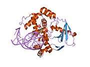
 structure of catalytically inactive q315a human 8-oxoguanine glycosylase complexed to 8-oxoguanine dna | |||||||||
| Identifiers | |||||||||
| Symbol | OGG_N | ||||||||
| Pfam | PF07934 | ||||||||
| Pfam clan | CL0407 | ||||||||
| InterPro | IPR012904 | ||||||||
| SCOP | 1ebm | ||||||||
| SUPERFAMILY | 1ebm | ||||||||
| |||||||||
8-Oxoguanine glycosylase also known as OGG1 is a DNA glycosylase enzyme that, in humans, is encoded by the OGG1 gene. It is involved in base excision repair. It is found in bacterial, archaeal and eukaryotic species.
Function
OGG1 is the primary enzyme responsible for the excision of 8-oxoguanine (8-oxoG), a mutagenic base byproduct that occurs as a result of exposure to reactive oxygen species (ROS). OGG1 is a bifunctional glycosylase, as it is able to both cleave the glycosidic bond of the mutagenic lesion and cause a strand break in the DNA backbone. Alternative splicing of the C-terminal region of this gene classifies splice variants into two major groups, type 1 and type 2, depending on the last exon of the sequence. Type 1 alternative splice variants end with exon 7 and type 2 end with exon 8. One set of spliced forms are designated 1a, 1b, 2a to 2e.[3] All variants have the N-terminal region in common. Many alternative splice variants for this gene have been described, but the full-length nature for every variant has not been determined. In eukaryotes, the N-terminus of this gene contains a mitochondrial targeting signal, essential for mitochondrial localization.[4] However, OGG1-1a also has a nuclear location signal at its C-terminal end that suppresses mitochondrial targeting and causes OGG1-1a to localize to the nucleus.[3] The main form of OGG1 that localizes to the mitochondria is OGG1-2a.[3] A conserved N-terminal domain contributes residues to the 8-oxoguanine binding pocket. This domain is organised into a single copy of a TBP-like fold.[5]
Despite the presumed importance of this enzyme, mice lacking Ogg1 have been generated and found to have a normal lifespan,[6] and Ogg1 knockout mice have a higher probability to develop cancer, whereas Mth1 gene disruption concomitantly suppresses lung cancer development in Ogg1-/- mice.[7] Interestingly, mice lacking Ogg1 have been shown to be prone to increased body weight and obesity, as well as high-fat diet induced insulin resistance.[8] There is some controversy as to whether deletion of Ogg1 actually leads to increased 8-oxo-dG levels: the HPLC-EC assay suggests up to 6 fold higher levels of 8-oxo-dG in nuclear DNA and 20-fold higher in mitochondrial DNA whereas the fappy-glycosylase assay indicates no change.
OGG1 deficiency and increased 8-oxo-dG in mice
_and_with_tumorigenesis_(B)._Brown_shows_8-oxo-dG.jpg)
Mice without a functional OGG1 gene have about a 5-fold increased level of 8-oxo-dG in their livers compared to mice with wild-type OGG1.[7] Mice defective in OGG1 also have an increased risk for cancer.[7] Kunisada et al.[10] irradiated mice without a functional OGG1 gene (OGG1 knock-out mice) and wild-type mice three times a week for forty weeks with UVB light at a relatively low dose (not enough to cause skin redness). Both types of mice had high levels of 8-oxo-dG in their epidermal cells three hours after irradiation. However, 24 hours later, the majority of 8-oxo-dG was absent from the epidermal cells of the wild-type mice but 8-oxo-dG remained elevated in the epidermal cells of the OGG1 knock-out mice. The irradiated OGG1 knock-out mice had more than twice the level of skin tumors compared to irradiated wild-type mice, and the rate of malignancy within the tumors was higher in the OGG1 knock-out mice (73%) than in the wild-type mice (50%).
As reviewed by Valavanidis et al.,[11] increased levels of 8-oxo-dG in a tissue can serve as a biomarker of oxidative stress. They also noted that increased levels of 8-oxo-dG are frequently found during carcinogenesis.
In the figure showing examples of mouse colonic epithelium, the colonic epithelium from a mouse on a normal diet was found to have a low level of 8-oxo-dG in its colonic crypts (panel A). However, a mouse likely undergoing colonic tumorigenesis (due to deoxycholate added to its diet[9]) was found to have a high level of 8-oxo-dG in its colonic epithelium (panel B). Deoxycholate increases intracellular production of reactive oxygen resulting in increased oxidative stress,[12][13] and this can lead to tumorigenesis and carcinogenesis.
Epigenetic control
In a breast cancer study, the methylation level of the OGG1 promoter was found to be anti-correlated with expression level of OGG1 messenger RNA.[14] This means that hypermethylation was associated with low expression of OGG1 and hypomethylation was correlated with over-expression of OGG1. Thus, OGG1 expression is under epigenetic control. Breast cancers with methylation levels of the OGG1 promoter that were more than two standard deviations either above or below the normal were each associated with reduced patient survival.[14]
OGG1 in cancers
OGG1 is the primary enzyme responsible for the excision of 8-oxo-2'-deoxyguanosine (8-oxo-dG). Even when OGG1 expression is normal, the presence of 8-oxo-dG is mutagenic since OGG1 is not 100% effective. Yasui et al.[15] examined the fate of 8-oxo-dG when this oxidized derivative of deoxyguanosine was inserted into a specific gene in 800 cells in culture. After replication of the cells, 8-oxo-dG was restored to G in 86% of the clones, probably reflecting accurate OGG1 base excision repair or translesion synthesis without mutation. G:C to T:A transversions occurred in 5.9% of the clones, single base deletions in 2.1% and G:C to C:G transversions in 1.2%. Together, these mutations were the most common, totalling 9.2% of the 14% of mutations generated at the site of the 8-oxo-dG insertion. Among the other mutations in the 800 clones analyzed, there were also 3 larger deletions, of sizes 6, 33 and 135 base pairs. Thus 8-oxo-dG can directly cause mutations, some of which may contribute to carcinogenesis.
If OGG1 expression is reduced in cells, increased mutagenesis, and therefore increased carcinogenesis would be expected. The table, below, lists cancers with reduced expression of OGG1.
| Cancer | Expression | Form of OGG1 | 8-oxo-dG | Evaluation method | Ref. |
|---|---|---|---|---|---|
| Head and neck cancer | Under-expression | OGG1-2a | - | messenger RNA | [16] |
| Adenocarcinoma of gastric cardia | Under-expression | cytoplasmic | increased | immunohistochemistry | [17] |
| Astrocytoma | Under-expression | total cell OGG1 | - | messenger RNA | [18] |
| Esophageal cancer | 48% Under-expression | nuclear | increased | immunohistochemistry | [19] |
| - | 40% Under-expression | cytoplasm | increased | immunohistochemistry | [19] |
OGG1 or OGG activity in blood, and cancer
OGG1 methylation levels in blood cells were measured in a prospective study of 582 US Veterans, median age 72, and followed for 13 years. High OGG1 methylation at a particular promoter region was associated with increased risk for any cancer, and in particular for risk of prostate cancer.[20]
Enzymatic activity excising 8-oxoguanine from DNA (OGG activity) was reduced in peripheral blood mononuclear cells (PBMCs), and in paired lung tissue, from patients with non–small cell lung cancer.[21] OGG activity was also reduced in PBMCs of patients with head and neck squamous cell carcinoma (HNSCC).[22]
Interactions
Oxoguanine glycosylase has been shown to interact with XRCC1[23] and PKC alpha.[24]
Pathology
References
- ↑ "Human PubMed Reference:".
- ↑ "Mouse PubMed Reference:".
- 1 2 3 Nishioka K, Ohtsubo T, Oda H, Fujiwara T, Kang D, Sugimachi K, Nakabeppu Y (1999). "Expression and differential intracellular localization of two major forms of human 8-oxoguanine DNA glycosylase encoded by alternatively spliced OGG1 mRNAs". Mol. Biol. Cell. 10 (5): 1637–52. doi:10.1091/mbc.10.5.1637. PMC 30487
 . PMID 10233168.
 - ↑ "Entrez Gene: OGG1 8-oxoguanine DNA glycosylase".
- ↑ Bjørås M, Seeberg E, Luna L, Pearl LH, Barrett TE (March 2002). "Reciprocal "flipping" underlies substrate recognition and catalytic activation by the human 8-oxo-guanine DNA glycosylase". J. Mol. Biol. 317 (2): 171–7. doi:10.1006/jmbi.2002.5400. PMID 11902834.
- ↑ Klungland A, Rosewell I, Hollenbach S, Larsen E, Daly G, Epe B, Seeberg E, Lindahl T, Barnes DE (November 1999). "Accumulation of premutagenic DNA lesions in mice defective in removal of oxidative base damage". Proc. Natl. Acad. Sci. U.S.A. 96 (23): 13300–5. doi:10.1073/pnas.96.23.13300. PMC 23942. PMID 10557315.
- 1 2 3 Sakumi K, Tominaga Y, Furuichi M, Xu P, Tsuzuki T, Sekiguchi M, Nakabeppu Y (2003). "Ogg1 knockout-associated lung tumorigenesis and its suppression by Mth1 gene disruption". Cancer Res. 63 (5): 902–5. PMID 12615700.
- ↑ Sampath H, Vartanian V, Rollins MR, Sakumi K, Nakabeppu Y, Lloyd RS (December 2012). "8-Oxoguanine DNA glycosylase (OGG1) deficiency increases susceptibility to obesity and metabolic dysfunction". PLoS ONE. 7 (12): e51697. doi:10.1371/journal.pone.0051697. PMC 3524114. PMID 23284747.
- 1 2 Prasad AR, Prasad S, Nguyen H, Facista A, Lewis C, Zaitlin B, Bernstein H, Bernstein C (2014). "Novel diet-related mouse model of colon cancer parallels human colon cancer". World J Gastrointest Oncol. 6 (7): 225–43. doi:10.4251/wjgo.v6.i7.225. PMC 4092339. PMID 25024814.
- ↑ Kunisada M, Sakumi K, Tominaga Y, Budiyanto A, Ueda M, Ichihashi M, Nakabeppu Y, Nishigori C (2005). "8-Oxoguanine formation induced by chronic UVB exposure makes Ogg1 knockout mice susceptible to skin carcinogenesis". Cancer Res. 65 (14): 6006–10. doi:10.1158/0008-5472.CAN-05-0724. PMID 16024598.
- ↑ Valavanidis A, Vlachogianni T, Fiotakis K, Loridas S (2013). "Pulmonary oxidative stress, inflammation and cancer: respirable particulate matter, fibrous dusts and ozone as major causes of lung carcinogenesis through reactive oxygen species mechanisms". Int J Environ Res Public Health. 10 (9): 3886–907. doi:10.3390/ijerph10093886. PMC 3799517. PMID 23985773.
- ↑ Tsuei J, Chau T, Mills D, Wan YJ. Bile acid dysregulation, gut dysbiosis, and gastrointestinal cancer. Exp Biol Med (Maywood). 2014 Nov;239(11):1489-504. doi: 10.1177/1535370214538743. PMID 24951470
- ↑ Ajouz H, Mukherji D, Shamseddine A. Secondary bile acids: an underrecognized cause of colon cancer. World J Surg Oncol. 2014 May 24;12:164. doi: 10.1186/1477-7819-12-164. Review. PMID 24884764
- 1 2 Fleischer T, Edvardsen H, Solvang HK, Daviaud C, Naume B, Børresen-Dale AL, Kristensen VN, Tost J (2014). "Integrated analysis of high-resolution DNA methylation profiles, gene expression, germline genotypes and clinical end points in breast cancer patients". Int. J. Cancer. 134 (11): 2615–25. doi:10.1002/ijc.28606. PMID 24395279.
- ↑ Yasui M, Kanemaru Y, Kamoshita N, Suzuki T, Arakawa T, Honma M (2014). "Tracing the fates of site-specifically introduced DNA adducts in the human genome". DNA Repair (Amst.). 15: 11–20. doi:10.1016/j.dnarep.2014.01.003. PMID 24559511.
- ↑ Mahjabeen I, Kayani MA (2016). "Loss of Mitochondrial Tumor Suppressor Genes Expression Is Associated with Unfavorable Clinical Outcome in Head and Neck Squamous Cell Carcinoma: Data from Retrospective Study". PLoS ONE. 11 (1): e0146948. doi:10.1371/journal.pone.0146948. PMC 4718451. PMID 26785117.
- ↑ Kohno Y, Yamamoto H, Hirahashi M, Kumagae Y, Nakamura M, Oki E, Oda Y (2016). "Reduced MUTYH, MTH1, and OGG1 expression and TP53 mutation in diffuse-type adenocarcinoma of gastric cardia". Hum. Pathol. 52: 145–52. doi:10.1016/j.humpath.2016.01.006. PMID 26980051.
- ↑ Jiang Z, Hu J, Li X, Jiang Y, Zhou W, Lu D (2006). "Expression analyses of 27 DNA repair genes in astrocytoma by TaqMan low-density array". Neurosci. Lett. 409 (2): 112–7. doi:10.1016/j.neulet.2006.09.038. PMID 17034947.
- 1 2 Kubo N, Morita M, Nakashima Y, Kitao H, Egashira A, Saeki H, Oki E, Kakeji Y, Oda Y, Maehara Y (2014). "Oxidative DNA damage in human esophageal cancer: clinicopathological analysis of 8-hydroxydeoxyguanosine and its repair enzyme". Dis. Esophagus. 27 (3): 285–93. doi:10.1111/dote.12107. PMID 23902537.
- ↑ Gao T, Joyce BT, Liu L, Zheng Y, Dai Q, Zhang Z, Zhang W, Shrubsole MJ, Tao MH, Schwartz J, Baccarelli A, Hou L (2016). "DNA methylation of oxidative stress genes and cancer risk in the Normative Aging Study". Am J Cancer Res. 6 (2): 553–61. PMC 4859680. PMID 27186424.
- ↑ Paz-Elizur T, Krupsky M, Blumenstein S, Elinger D, Schechtman E, Livneh Z (2003). "DNA repair activity for oxidative damage and risk of lung cancer". J. Natl. Cancer Inst. 95 (17): 1312–9. doi:10.1093/jnci/djg033. PMID 12953085.
- ↑ Paz-Elizur T, Ben-Yosef R, Elinger D, Vexler A, Krupsky M, Berrebi A, Shani A, Schechtman E, Freedman L, Livneh Z (2006). "Reduced repair of the oxidative 8-oxoguanine DNA damage and risk of head and neck cancer". Cancer Res. 66 (24): 11683–9. doi:10.1158/0008-5472.CAN-06-2294. PMID 17178863.
- ↑ Marsin S, Vidal AE, Sossou M, Ménissier-de Murcia J, Le Page F, Boiteux S, de Murcia G, Radicella JP (November 2003). "Role of XRCC1 in the coordination and stimulation of oxidative DNA damage repair initiated by the DNA glycosylase hOGG1". J. Biol. Chem. 278 (45): 44068–74. doi:10.1074/jbc.M306160200. PMID 12933815.
- ↑ Dantzer F, Luna L, Bjørås M, Seeberg E (June 2002). "Human OGG1 undergoes serine phosphorylation and associates with the nuclear matrix and mitotic chromatin in vivo". Nucleic Acids Res. 30 (11): 2349–57. doi:10.1093/nar/30.11.2349. PMC 117190. PMID 12034821.
- ↑ Osorio A, Milne RL, Kuchenbaecker K, Vaclová T, Pita G, Alonso R, Peterlongo P, Blanco I, de la Hoya M, Duran M, Díez O, Ramón Y, Cajal T, Konstantopoulou I, Martínez-Bouzas C, Andrés Conejero R, Soucy P, McGuffog L, Barrowdale D, Lee A, Arver B, Rantala J, Loman N, Ehrencrona H, Olopade OI, Beattie MS, Domchek SM, Nathanson K, Rebbeck TR, Arun BK, Karlan BY, Walsh C, Lester J, John EM, Whittemore AS, Daly MB, Southey M, Hopper J, Terry MB, Buys SS, Janavicius R, Dorfling CM, van Rensburg EJ, Steele L, Neuhausen SL, Ding YC, Hansen TV, Jønson L, Ejlertsen B, Gerdes AM, Infante M, Herráez B, Moreno LT, Weitzel JN, Herzog J, Weeman K, Manoukian S, Peissel B, Zaffaroni D, Scuvera G, Bonanni B, Mariette F, Volorio S, Viel A, Varesco L, Papi L, Ottini L, Tibiletti MG, Radice P, Yannoukakos D, Garber J, Ellis S, Frost D, Platte R, Fineberg E, Evans G, Lalloo F, Izatt L, Eeles R, Adlard J, Davidson R, Cole T, Eccles D, Cook J, Hodgson S, Brewer C, Tischkowitz M, Douglas F, Porteous M, Side L, Walker L, Morrison P, Donaldson A, Kennedy J, Foo C, Godwin AK, Schmutzler RK, Wappenschmidt B, Rhiem K, Engel C, Meindl A, Ditsch N, Arnold N, Plendl HJ, Niederacher D, Sutter C, Wang-Gohrke S, Steinemann D, Preisler-Adams S, Kast K, Varon-Mateeva R, Gehrig A, Stoppa-Lyonnet D, Sinilnikova OM, Mazoyer S, Damiola F, Poppe B, Claes K, Piedmonte M, Tucker K, Backes F, Rodríguez G, Brewster W, Wakeley K, Rutherford T, Caldés T, Nevanlinna H, Aittomäki K, Rookus MA, van Os TA, van der Kolk L, de Lange JL, Meijers-Heijboer HE, van der Hout AH, van Asperen CJ, Gómez Garcia EB, Hoogerbrugge N, Collée JM, van Deurzen CH, van der Luijt RB, Devilee P, Olah E, Lázaro C, Teulé A, Menéndez M, Jakubowska A, Cybulski C, Gronwald J, Lubinski J, Durda K, Jaworska-Bieniek K, Johannsson OT, Maugard C, Montagna M, Tognazzo S, Teixeira MR, Healey S, Investigators K, Olswold C, Guidugli L, Lindor N, Slager S, Szabo CI, Vijai J, Robson M, Kauff N, Zhang L, Rau-Murthy R, Fink-Retter A, Singer CF, Rappaport C, Geschwantler Kaulich D, Pfeiler G, Tea MK, Berger A, Phelan CM, Greene MH, Mai PL, Lejbkowicz F, Andrulis I, Mulligan AM, Glendon G, Toland AE, Bojesen A, Pedersen IS, Sunde L, Thomassen M, Kruse TA, Jensen UB, Friedman E, Laitman Y, Shimon SP, Simard J, Easton DF, Offit K, Couch FJ, Chenevix-Trench G, Antoniou AC, Benitez J (2014). "DNA glycosylases involved in base excision repair may be associated with cancer risk in BRCA1 and BRCA2 mutation carriers". PLoS Genet. 10 (4): e1004256. doi:10.1371/journal.pgen.1004256. PMC 3974638. PMID 24698998.
Further reading
- Boiteux S, Radicella JP (2000). "The human OGG1 gene: structure, functions, and its implication in the process of carcinogenesis". Arch. Biochem. Biophys. 377 (1): 1–8. doi:10.1006/abbi.2000.1773. PMID 10775435.
- Park J, Chen L, Tockman MS, Elahi A, Lazarus P (2004). "The human 8-oxoguanine DNA N-glycosylase 1 (hOGG1) DNA repair enzyme and its association with lung cancer risk". Pharmacogenetics. 14 (2): 103–9. doi:10.1097/00008571-200402000-00004. PMID 15077011.
- Hung RJ, Hall J, Brennan P, Boffetta P (2005). "Genetic polymorphisms in the base excision repair pathway and cancer risk: a HuGE review". Am. J. Epidemiol. 162 (10): 925–42. doi:10.1093/aje/kwi318. PMID 16221808.
- Mirbahai L, Kershaw RM, Green RM, Hayden RE, Meldrum RA, Hodges NJ (2010). "Use of a molecular beacon to track the activity of base excision repair protein OGG1 in live cells". DNA Repair (Amst.). 9 (2): 144–52. doi:10.1016/j.dnarep.2009.11.009. PMID 20042377.
External links
- oxoguanine glycosylase 1, human at the US National Library of Medicine Medical Subject Headings (MeSH)
PDB gallery | ||||||||||||||||||||||||||||||||||||||||||||||||||
|---|---|---|---|---|---|---|---|---|---|---|---|---|---|---|---|---|---|---|---|---|---|---|---|---|---|---|---|---|---|---|---|---|---|---|---|---|---|---|---|---|---|---|---|---|---|---|---|---|---|---|
| ||||||||||||||||||||||||||||||||||||||||||||||||||


This article incorporates text from the public domain Pfam and InterPro IPR012904