Optic radiation
| Optic radiation | |
|---|---|
 Colour-coded diagram showing radiations in quadrants from retinal disc through the brain | |
| Details | |
| Identifiers | |
| Latin | radiatio optica |
| NeuroNames | ancil-529 |
| TA |
A14.1.08.673 A14.1.09.542 A14.1.09.547 |
| FMA | 61941 |
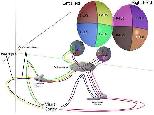
The optic radiation (also known as the geniculocalcarine tract, the geniculostriate pathway, and posterior thalamic radiation) are axons from the neurons in the lateral geniculate nucleus to the primary visual cortex. The optic radiation receives blood through deep branches of the middle cerebral artery and posterior cerebral artery.
They carry visual information through two divisions (called upper and lower division) to the visual cortex (also called striate cortex) along the calcarine fissure. There is one such tract on each side of the brain. If a lesion only exists in one optic radiation, the consequence is called quadrantanopia, which implies that only the respective superior or inferior quadrant of the visual field is affected.
Structure
The upper division:
- Projects to the upper bank of the calcarine fissure, called the cuneus
- Contains input from the superior retinal quadrants, which represents the inferior visual field quadrants
- Transection causes contralateral lower quadrantanopia
- Lesions that involve both cunei cause a lower altitudinal hemianopia (altitudinopia)
The lower division:
- Loops from the lateral geniculate body anteriorly (Meyer's loop), then posteriorly, to terminate in the lower bank of the calcarine sulcus, called the lingual gyrus
- Contains input from the inferior retinal quadrants, which represents the superior visual field quadrants
- Transection causes contralateral upper quadrantanopia
- Transection of both lingual gyri causes an upper altitudinal hemianopia
Parts
A distinctive feature of the optic radiations is that they split into two parts on each side:
| Source | Path | Information | Damage |
| Fibers from the inferior retina (also called "Meyer's loop" or "Archambault's loop") | must pass through the temporal lobe by looping around the inferior horn of the lateral ventricle. | Carry information from the superior part of the visual field | A lesion in the temporal lobe that results in damage to Meyer's loop causes a characteristic loss of vision in a superior quadrant (quadrantanopia or "pie in the sky" defect.) |
| Fibers from the superior retina (also called "Baum's loop") | travel straight back through the parietal lobe to the occipital lobe in the retrolenticular limb of the internal capsule to the visual cortex. | Carry information from the inferior part of the visual field | Taking the shorter path, these fibers are less susceptible to damage. Damage caused is characteristically called "Pie in the floor" defect or inferior quadrantanopia. |

Function
The optic radiation contains tracts which transmit visual information from the retina of the eye to the visual cortex. Lesions of the optic radiations are usually unilateral and commonly vascular in origin. Field defects therefore develop abruptly, in contrast to the slow progression of defects associated with tumors.
Clinical significance
Examination
Tracts contained within the optic radiation are examined as part of a cranial nerve examination.
Additional images
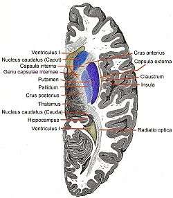 Horizontal section of right cerebral hemisphere.
 Diagram of the tracts in the internal capsule.
- 3D schematic representation of optic tracts
External links
| Wikimedia Commons has media related to Optic radiation. |
- Kier LE, Staib LH, Davis LM, Bronen RA (1 May 2004). "MR Imaging of the Temporal Stem: Anatomic Dissection Tractography of the Uncinate Fasciculus, Inferior Occipitofrontal Fasciculus, and Meyer's Loop of the Optic Radiation". Am J Neuroradiol. 25 (5): 677–691. PMID 15140705. Retrieved 2007-12-19.
- http://www2.umdnj.edu/~neuro/studyaid/Practical2000/Q34.htm
- A 3D model of optic tract and optic radiation
| Related | ||
|---|---|---|