Nanoscale secondary ion mass spectrometry
Nanoscale secondary ion mass spectrometry (nanoSIMS or nano secondary ion mass spectrometry) is a nanoscopic scale resolution chemical imaging mass spectrometer based on secondary ion mass spectrometry.[1] It works based on a coaxial optical design of the ion gun and the secondary ion extraction, and on an original magnetic sector mass spectrometer with multicollection.[2]
NanoSIMS not only refers to the technique used, but also the mass spectrometer specialized for this method. There are currently only 22 NanoSIMS in the world.[3]
How it works
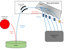
The magnetic sector mass spectrometer causes a physical separation of ions of a different mass-to-charge ratio. The physical separation of the secondary ions is caused by the Lorentz force when the ions pass through a magnetic field that is perpendicular to the velocity vector of the secondary ions. The Lorentz force states that a particle will experience a force
![\mathbf {F} =q\left[\mathbf {E} +(\mathbf {v} \times \mathbf {B} )\right]](../I/m/f0139e901d2aa4f20d3deb80a4e6dd56cd3321a6.svg)
when it maintains a charge q and travels through an electric field E and magnetic field B with a velocity v. The secondary ions that leave the surface of the sample typically have a kinetic energy of a few electron volts (eV), although a rather small portion have been found to have energy of a few keV. An electrostatic field captures the secondary ions that leave the sample surface; these extracted ions are then transferred to a mass spectrometer. In order to achieve precise isotope measurements, there is a need for high transmission and high mass resolution. High transmission refers to the low loss of secondary ions between the sample surface and the detector, and high mass resolution refers to the ability to efficiently separate the secondary ions (or molecules of interest) from other ions and/or ions of similar mass. Primary ions will collide with the surface at a specific frequency per unit of surface area. The collision that occurs causes atoms to sputter from the sample surface, and of these atoms only a small amount will undergo ionization. These become secondary ions, which are then detected after transfer through the mass spectrometer. Each primary ion generates a number of secondary ions of an isotope that will reach the detector to be counted. The count rate is determined by

where I(iM)is the count rate of the isotope iM of element M. The counting rate of the isotope is dependent on the concentration, XM and the element's isotopic abundance, denoted Ai. Because the primary ion beam determines the secondary ions, Y, that are sputtered, the density of the primary ion beam, db, which is defined as the amount of ions per second per unit of surface area, will affect a portion of the surface area of the sample, S, with an even distribution of the primary ions. Of the sputtered secondary ions, there is only a fraction that will be ionized, Yi. The probability that any ion will be successfully transferred from mass spectrometer to detector is T. The product of Yi and T determines the amount of isotopes that will be ionized, as well as detected, so it is considered the useful yield. [4]
Instrumentation
The NanoSIMS 50L is the SIMS microprobe for isotopic and trace element analysis at high spatial resolution. Original design of the instrument was conceived by Georges Slodzian at the University of Paris Sud in France.[5]
The mechanism of nanoSIMS is based on secondary ion mass spectrometry. This instrument can characterize the nanostructured materials with complex composition that are increasingly important candidates for energy generation and storage.
NanoSIMS is able to create nanoscale maps of elemental composition, parallel acquisition of seven masses, isotopic identification, combining the high mass resolution, subparts-per-million sensitivity of conventional SIMS with spatial resolution down to 50 nm and fast acquisition (DC mode, not pulsed).[6]
Applications
NanoSIMS combined with fluorescence microscopy can be used as a tool for subcellular imaging of isotopically labeled platinum-based anticancer drugs.[7]
It also allows precise isotopic and elemental measurements of deep sub-micron areas, grains or inclusions from the different geological and spatial samples.[8]
NanoSIMS has also proved useful in studying cosmochemical issues, where samples that were studied included sections of meteorites, single, micro- or sub-micrometer-sized grains, such as presolar grains distributed on gold foils, as well as microtome sections or those that were prepared by the focused ion beam (FIB) technique. NanoSIMS can be combined with transmission electron microscopy (TEM) when using microtome or FIB sections. This combination allows for correlated mineralogical and isotopic studies in situ at a sub-micrometer scale. Being able to study presolar grains includes presolar silicates, presolar oxides, as well as presolar silicon carbide (SiC) and graphite grains.
In the field of biology it proved useful in analyzing an antigen bound to an antibody that had been immobilized for analysis. In one study it was found to be a label-free method, allowing for localization and quantitative analysis of antigen-antibody binding on a surface, where nanoSIMS was chosen as one method in order to obtain imaging on an atomic level of the surface that was used in the binding.[9] In microbiology, there are more opportunities with nanoSIMS. It opened up the possibility for coupling phylogenetic identity and metabolic function in mixed microbial communities of single cells.[10] The high resolution that it offers allows intracellular measurement of accumulations and fluxes of molecules containing various stable isotopes.[11]
It is particularly useful in materials research because of its high sensitivity at high mass resolution, which allow for trace element imaging and quatification.[12]
References
- ↑ Herrmann, Anke M.; Ritz, Karl; Nunan, Naoise; Clode, Peta L.; Pett-Ridge, Jennifer; Kilburn, Matt R.; Murphy, Daniel V.; O’Donnell, Anthony G.; Stockdale, Elizabeth A. (2007). "Nano-scale secondary ion mass spectrometry — A new analytical tool in biogeochemistry and soil ecology: A review article". Soil Biology and Biochemistry. 39 (8): 1835–1850. doi:10.1016/j.soilbio.2007.03.011. ISSN 0038-0717.
- ↑ Oxford University,Department of Materials
- ↑ Stanford Nano Center
- ↑ Hoppe, Peter; Cohen, Stephanie; Meibom, Anders (2013). "NanoSIMS: Technical Aspects and Applications in Cosmochemistry and Biological Geochemistry". Geostandards and Geoanalytical Research. 37 (2): 111–154. doi:10.1111/j.1751-908X.2013.00239.x.
- ↑ "CAMECA NanoSIMS: High Resolution Ion Microprobe for Ultra Fine Feature Analysis". www.cameca.com. Retrieved April 20, 2016.
- ↑ Cameca NanoSIMS 50L
- ↑ Legin, Anton A.; Schintlmeister, Arno; Jakupec, Michael A.; Galanski, Markus; Lichtscheidl, Irene; Wagner, Michael; Keppler, Bernhard K. (2014). "NanoSIMS combined with fluorescence microscopy as a tool for subcellular imaging of isotopically labeled platinum-based anticancer drugs". Chemical Science. 5 (8): 3135. doi:10.1039/c3sc53426j. ISSN 2041-6520.
- ↑ J. Moreau et al., SCIENCE.
- ↑ Dauphas, Stéphanie; Delhaye, Thomas; Lavastre, Olivier; Corlu, Anne; Guguen-Guillouzo, Christiane; Ababou-Girard, Soraya; Geneste, Florence (2008). "Localization and Quantitative Analysis of Antigen−Antibody Binding on 2D Substrate Using Imaging NanoSIMS". Analytical Chemistry. 80 (15): 5958–5962. doi:10.1021/ac800602q. ISSN 0003-2700.
- ↑ Musat, N.; Halm, H.; Winterholler, B.; Hoppe, P.; Peduzzi, S.; Hillion, F.; Horreard, F.; Amann, R.; Jorgensen, B. B.; Kuypers, M. M. M. (2008). "A single-cell view on the ecophysiology of anaerobic phototrophic bacteria". Proceedings of the National Academy of Sciences. 105 (46): 17861–17866. doi:10.1073/pnas.0809329105. ISSN 0027-8424.
- ↑ "Application of the CAMECA NanoSIMS : Cell Biology". www.cameca.com.
- ↑ "CAMECA NanoSIMS Application to Materials Research: Segregation & Diffusion in Polycrystalline". www.cameca.com.