Mitomycins

The mitomycins are a family of aziridine-containing natural products isolated from Streptomyces caespitosus or Streptomyces lavendulae.[1] They include mitomycin A, mitomycin B, and mitomycin C. When the name mitomycin occurs alone, it usually refers to mitomycin C; it is the international nonproprietary name for mitomycin C.
Biosynthesis
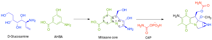
In general, the biosynthesis of all mitomycins[2] proceeds via combination of 3-amino-5-hydroxybenzoic acid (AHBA), D-glucosamine, and carbamoyl phosphate, to form the mitosane core, followed by specific tailoring steps. The key intermediate, AHBA, is a common precursor to other anticancer drugs, such as rifamycin and ansamycin.
Specifically, the biosynthesis begins with the addition of phosphoenolpyruvate (PEP) to erythrose-4-phosphate (E4P) with a yet undiscovered enzyme, which is then ammoniated to give 4-amino-3-deoxy-D-arabino heptulosonic acid-7-phosphate (aminoDHAP). Next, DHQ synthase catalyzes a ring closure to give 4-amino3-dehydroquinate (aminoDHQ), which is then undergoes a double oxidation via aminoDHQ dehydratase to give 4-amino-dehydroshikimate (aminoDHS). The key intermediate, 3-amino-5-hydroxybenzoic acid (AHBA), is made via aromatization by AHBA synthase.

Synthesis of the key intermediate, 3-amino-5-hydroxy-benzoic acid.
The mitosane core is synthesized as shown below via condensation of AHBA and D-glucosamine, although no specific enzyme has been characterized that mediates this transformation. Once this condensation has occurred, the mitosane core is tailored by a variety of enzymes. Both the sequence and the identity of these steps are yet to be determined.
- Complete reduction of C-6 - Likely via F420-dependent tetrahydromethanopterin (H4MPT) reductase and H4MPT:CoM methyltransferase
- Hydroxylation of C-5, C-7 (followed by transamination), and C-9a. - Likely via cytochrome P450 monooxygenase or benzoate hydroxylase
- O-Methylation at C-9a - Likely via SAM dependent methyltransferase
- Oxidation at C-5 and C8 - Unknown
- Intramolecular amination to form aziridine - Unknown
- Carbamoylation at C-10 - Carbamoyl transferrase, with carbamoyl phosphate (C4P) being derived from L-citrulline or L-arginine
Biological effects
In the bacterium Legionella pneumophila, mitomycin C induces competence for transformation.[3] Natural transformation is a process of DNA transfer between cells, and is regarded as a form of bacterial sexual interaction. In the fruit fly Drosophila melanogaster, exposure to mitomycin C increases recombination during meiosis, a key stage of the sexual cycle.[4] In the plant Arabidopsis thaliana, mutant strains defective in genes necessary for recombination during meiosis and mitosis are hypersensitive to killing by mitomycin C.[5] It has been suggested that these, and other related findings, can be explained by the idea that during sexual processes in prokaryotes (transformation) and eukaryotes (meiosis) DNA crosslinks and other damages introduced by mitomycin C are removed by recombinational repair.[6]
Mitomycin C has recently been found to have very good activity against stationary phase[7] and against persisters[8] created by Borrelia burgdorferi, the causative agent of lyme disease.
References
- ↑ Danshiitsoodol N, de Pinho CA, Matoba Y, Kumagai T, Sugiyama M (2006). "The mitomycin C (MMC)-binding protein from MMC-producing microorganisms protects from the lethal effect of bleomycin: crystallographic analysis to elucidate the binding mode of the antibiotic to the protein". J Molec Biol. 360 (2): 398–408. doi:10.1016/j.jmb.2006.05.017. PMID 16756991.
- ↑ Mao Y.; Varoglu M.; Sherman D.H. (April 1999). "Molecular characterization and analysis of the biosynthetic gene cluster for the antitumor antibiotic mitomycin C from Streptomyces Iavendulae NRRL 2564.". Chemistry and Biology. 6 (4): 251–263. doi:10.1016/S1074-5521(99)80040-4. PMID 10099135.
- ↑ Charpentier X, Kay E, Schneider D, Shuman HA (March 2011). "Antibiotics and UV radiation induce competence for natural transformation in Legionella pneumophila". J. Bacteriol. 193 (5): 1114–21. doi:10.1128/JB.01146-10. PMC 3067580
 . PMID 21169481.
 - ↑ Schewe MJ, Suzuki DT, Erasmus U (July 1971). "The genetic effects of mitomycin C in Drosophila melanogaster. II. Induced meiotic recombination". Mutat. Res. 12 (3): 269–79. doi:10.1016/0027-5107(71)90015-7. PMID 5563942.
- ↑ Bleuyard JY, Gallego ME, Savigny F, White CI (February 2005). "Differing requirements for the Arabidopsis Rad51 paralogs in meiosis and DNA repair". Plant J. 41 (4): 533–45. doi:10.1111/j.1365-313X.2004.02318.x. PMID 15686518.
- ↑ Bernstein H, Bernstein C, Michod RE (2012). DNA repair as the primary adaptive function of sex in bacteria and eukaryotes. Chapter 1: pp.1-49 in: DNA Repair: New Research, Sakura Kimura and Sora Shimizu editors. Nova Sci. Publ., Hauppauge, N.Y. ISBN 978-1-62100-808-8 https://www.novapublishers.com/catalog/product_info.php?products_id=31918
- ↑ Feng, Jie; Shi, Wanliang; Zhang, Shuo; Zhang, Ying (3 June 2015). "Identification of new compounds with high activity against stationary phase Borrelia burgdorferi from the NCI compound collection". Emerging Microbes & Infections. 4 (5): e31. doi:10.1038/emi.2015.31.
- ↑ Sharma, Bijaya; Brown, Autumn V.; Matluck, Nicole E.; Hu, Linden T.; Lewis, Kim (26 May 2015). "Borrelia burgdorferi, the Causative Agent of Lyme Disease, Forms Drug-Tolerant Persister Cells". Antimicrobial Agents and Chemotherapy: AAC.00864–15. doi:10.1128/AAC.00864-15.
Further reading
- Hata, T.; Sano, Y.; Sugawara, R.; Matsumae, A.; Kanamori, K.; Shima, T.; Hoshi, T. (1956). "Mitomycin, a new antibiotic from Streptomyces.". J. Antibiot. Ser. A. 9: 141–146.
- Fukuyama, T.; Yang, L. "Total Synthesis of (±)-Mitomycins via Isomitomycin A". J. Am. Chem. Soc. '1987'. 109: 7881–7882. doi:10.1021/ja00259a046.
- Mao, Y.; Varoglu, M.; Sherman, D.H. (April 1999). "Molecular characterization and analysis of the biosynthetic cluster for the antitumor antibiotic mitomycin C from Streptomyces lavendulae NRRL 2564". Chemistry & Biology. 6 (4): 251–263. doi:10.1016/S1074-5521(99)80040-4. PMID 10099135.
- Varoglu, M.; Mao, Y.; Sherman, D.H. (2001). "Mapping the Biosynthetic Pathway by Functional Analysis of the MitM Aziridine N-Methyltransferase". J. Am. Chem. Soc. 123: 6712–6713. doi:10.1021/ja015646l. and references therein.