Marginal zone
| Marginal zone | |
|---|---|
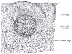 Transverse section of a portion of the spleen. | |
| Identifiers | |
| FMA | 15852 |
The marginal zone is the region at the interface between the non-lymphoid red pulp and the lymphoid white-pulp of the spleen. (Some sources consider it to be the part of red pulp which borders on the white pulp, while other sources consider it to be neither red pulp nor white pulp.)
A marginal zone also exists in lymph nodes.[1]
Composition and markers
It is composed of cells derived primarily from the myeloid compartment of bone marrow differentiation. More recently, a population of neutrophils has been described to populate peripheral areas of the marginal zone.[2] At least three distinct cellular markers can be used to identify cells of the marginal zone, MOMA-1, ERTR-9 and MARCO.
Function
The major role of marginal zone is to trap particulate antigen from the circulation and present the antigen to the lymphocytes of the spleen.
Experiments have shown that inert latex beads as well as live bacteria such as Escherichia coli and Listeria monocytogenes are trapped by the marginal zone. However, only immunogenic substances, i.e. bacteria, are trafficked to the T and B cell zones of the white-pulp and are efficiently presented to elicit an immune response.
Marginal Zone Circulation
The marginal zone (MZ) is a highly transited area that receives large amounts of blood from the general circulation. Remarkably, the splenic microvasculature shows striking differences in mice and humans. In humans, the spleen receives blood from the splenic artery, which branches into central and penicillary arterioles .[3] Owing to the absence of a histologically defined marginal sinus, the blood flowing in penicillary arterioles directly drains into capillaries of the red pulp and perifollicular zone. The perifollicular zone is a well-defined area of decreased resistance that separates the MZ from the red pulp. Both the perifollicular zone and the red pulp consist of an open circulatory system of blood-filled spaces known as splenic cords, which have no defined endothelial delimitation and are in close contact with the venous sinusoidal vessels of the red pulp.[4]
Lymphocytes
Marginal zone lymphocytes are a type of B cell (Marginal-zone B cell, abbreviated "MZ B cell") created there, capable of binding IgM-antigen complexes. They are notable for their ability to serve several different roles in the immune system. MZ B cells express polyreactive BCRs that bind to multiple microbial molecular patterns.[5]
See also
References
- ↑ http://www.pathologyoutlines.com/lymphnodes.html[]
- ↑ Puga, Irene; Cols, Montserrat; Barra, Carolina M; He, Bing; Cassis, Linda; Gentile, Maurizio; Chen, Kang; Cerutti, Andrea (December 2011). "B cell–helper neutrophils stimulate the diversification and production of immunoglobulin in the marginal zone of the spleen". Nature Immunology. 13 (2): 170–80. doi:10.1038/ni.2194. PMID 22197976.
- ↑ Mebius, Reina E.; Kraal, Georg (August 2005). "Structure and function of the spleen". Nature Reviews Immunology. 5 (1): 606–616. doi:10.1038/nri1669. PMID 16056254. Retrieved 30 April 2015.
- ↑ Steiniger, Birte; Timphus,, Eva Maria; Barth, Peter J. (December 2006). "The splenic marginal zone in humans and rodents: an enigmatic compartment and its inhabitants". Histochemistry and Cell Biology. 126 (6): 641–648. doi:10.1007/s00418-006-0210-5. PMID 16816939.
- ↑ Martin, Flavius; Kearney, John F. (May 2002). "Marginal-zone B cells". Nature Reviews Immunology. 2 (5): 323–35. doi:10.1038/nri799. PMID 12033738.
External links
- Pillai S, Cariappa A, Moran ST (2005). "Marginal zone B cells". Annual Review of Immunology. 23: 161–96. doi:10.1146/annurev.immunol.23.021704.115728. PMID 15771569.
- Ferguson AR, Corley RB (2005). "Accumulation of marginal zone B cells and accelerated loss of follicular dendritic cells in NF-κB p50-deficient mice". BMC Immunology. 6: 8. doi:10.1186/1471-2172-6-8. PMC 1087843
 . PMID 15836790.
 - Marginal zone lymphomas
- Song H, Cerny J (December 2003). "Functional heterogeneity of marginal zone B cells revealed by their ability to generate both early antibody-forming cells and germinal centers with hypermutation and memory in response to a T-dependent antigen". The Journal of Experimental Medicine. 198 (12): 1923–35. doi:10.1084/jem.20031498. PMC 2194154. PMID 14662910.
- Kraal G (1992). "Cells in the marginal zone of the spleen". International Review of Cytology. 132: 31–74. doi:10.1016/S0074-7696(08)62453-5. PMID 1555921.
- Kumararatne DS, MacLennan IC (November 1981). "Cells of the marginal zone of the spleen are lymphocytes derived from recirculating precursors". European Journal of Immunology. 11 (11): 865–9. doi:10.1002/eji.1830111104. PMID 6976895.