Sensory root of ciliary ganglion
| Sensory root of ciliary ganglion | |
|---|---|
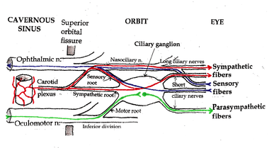 Pathways in the Ciliary Ganglion. Green = parasympathetic; Red = sympathetic; Blue = sensory | |
| Details | |
| From | nasociliary nerve |
| Identifiers | |
| Latin | ramus sensoria ganglii ciliaris |
| TA | A14.2.01.026 |
| FMA | 52672 |
Sensory fibers from the eyeball (the cornea, iris, and ciliary body) run posteriorly through the short ciliary nerves and pass through the ciliary ganglion without forming synapses. They leave the ciliary ganglion in the sensory root of ciliary ganglion, which joins the nasociliary nerve -- a branch of the ophthalmic nerve. From there, the signal travels back through the ophthalmic nerve to the trigeminal nerve and back into specific nuclei in the thalamus where they are relayed to areas in the cerebral cortex.
Variability
The exact distribution of sensory fibers, like the distribution of sympathetic fibers, is anatomically variable. There are alternate pathways to the eye for both sympathetic and sensory fibers, and the precise anatomy varies from person to person. Since the result is the same regardless of how the fibers reach the eye, the presence of sympathetic and sensory fibers in the ciliary ganglion (the contributions of the “sensory” and “sympathetic” roots) is of no functional significance.