Loa loa
| Loa loa | |
|---|---|
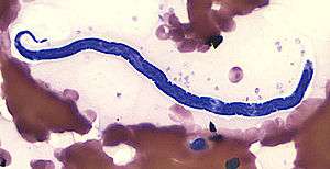 | |
| Loa loa microfilaria in thin blood smear (Giemsa stain) | |
| Scientific classification | |
| Kingdom: | Animalia |
| Phylum: | Nematoda |
| Class: | Chromadorea |
| Order: | Spirurida |
| Superfamily: | Filarioidea |
| Family: | Onchocercidae |
| Genus: | Loa |
| Species: | L. loa |
| Binomial name | |
| Loa loa (Cobbold, 1864) | |
| Synonyms | |
|
Filaria loa Cobbold, 1864 | |
Loa loa is the filarial nematode (roundworm) species that causes Loa loa filariasis. It is commonly known as the "eye worm". Loa Loa is commonly found in Africa and India.[1] It mainly inhabits rain forests in West Africa and has native origins in Ethiopia.[2]
L. loa is one of three parasitic filarial nematodes that cause subcutaneous filariasis in humans. The other two are Mansonella streptocerca and Onchocerca volvulus (causes river blindness).
Maturing larvae and adults of the "eye worm" occupy the subcutaneous layer of the skin – the fat layer – of humans, causing disease. The young larvae develop in horseflies of the genus Chrysops (deer flies, yellow flies), including the species C. dimidiata and C. silacea, which infect humans by biting them.
Biology
Morphology
Loa loa worms have a simple body consisting of a head, body, and tail. Males range from 20mm to 34mm long and 350μm to 430μm wide. Females range from 20mm to 70mm long and can be about 425μm wide. They vary in color.[1]
Life cycle [3]
The human remains the definitive host where the parasitic worm attains sexual maturity and the fly species serve as intermediate hosts needed for the morphological development of the worm and microfilariae production.
Two species of Chrysops deerflies, C. silacea and C. dimidiata are the two vectors for Loa loa filariasis disease.
- An infected fly introduces third-stage filarial larvae into the skin by penetrating into the bite wound.
- The larvae grow into adults which commonly live in subcutaneous tissue.
- The adults produce microfilariae that measure about 250 to 300 micrometers in length and six to eight micrometers wide. Microfilariae tend to reside are more likely found within spinal fluids, urine, and sputum. During daytime, the microfilariae can be found in the circulating bloodstream. However, while in the non-circulation phase, microfilariae can be found in the lungs. Female worms measure about 40 to 70 millimeters in length and 0.5 millimeters in diameter. Male worms measure about 30 to 34 millimeters in length and 0.35 to 0.43 millimeters in diameter.
- The microfilariae get ingested by a fly during a blood meal.
- Once it is inside the host, the microfilariae lose its sheaths and goes to the fly’s midgut through the hemocoel to the thoracic muscles of the arthropod.
- Once it does that, the microfilariae develop into first-stage larvae.
- Afterwards, it develops into third-stage infectious larvae.
- At the third-stage larvae, it migrates to the fly’s proboscis.
- Then, it can infect another human when the fly takes another blood meal and the cycle repeats.
Disease
Pathogenesis
Loa loa parasites infect human hosts by travelling from the entry site through subcutaneous tissues and causing inflammation in the skin wherever they travel. If a parasite stops in one place for a short period of time, the human host will suffer from local inflammation known as Calabar swellings. These are localized, tense, inflammatory pruritic subcutaneous edema seen in joints of extremities, lasting for 1–3 days. They represent areas of angioedema resulting from a host response to allergens released by the maturating worm and its metabolic products.[4] Calabar swellings often occur in the wrist and ankle joints but disappear as soon as the parasite begins to move again. Parasites can also travel through and infect the eye, causing the swelling of the eye. Common symptoms include itching, joint pain, fatigue, and death.[1]
Diagnosis and treatment
The main methods of diagnosis include the presence of microfilariae in the blood, the presence of a worm in the eye, and the presence of skin swellings. Surgical removal of the worm can easily be performed. The common treatment for the disease is the use of the drug Ivermectin.[1]
Ivermectin has become the most common antiparasitic agent used worldwide but can lead to residual microfilarial load when given in the management of loiasis. High microfilarial loads should be decreased by a course of Ivermectin, a prolonged administration of albendazole, or cytapheresis sessions to prevent occurrence of serious adverse events, including fatal encephalopathy induced by dying microfilariae. Cytapheresis is helpful in decreasing very high microfilarial loads up to 75%. Diethylcarbamazine kills both microfilariae and adult worms but has more severe side effects and can be fatal.
In the cause of treatment for a female patient in Lagos, Nigeria, patient complained of increase pain and swelling in her left palm, which progressed to temporary immobilization of the palm i.e. patient complained she couldn't use the palm especially in driving. After the 21-day treatment patient identified a small swelling in the left area of her lower abdomen. Swelling was itchy and revealed a dead worm. Swelling disappeared and healed after worm was removed without any major treatment besides cleaning of the opening.
References
- 1 2 3 4 Schmidt, Gerald et al. "Foundations of Parasitology". 7th ed. McGraw Hill, New York, NY, 2005.
- ↑ Thomson, MC; Obsomer, V; Dunne, M; Connor, SJ; Molyneux, DH. "Satellite mapping of Loa loa prevalence in relation to ivermectin use in west and central Africa". The Lancet. 356 (9235): 1077–1078. doi:10.1016/s0140-6736(00)02733-1.
- ↑ Prevention, CDC - Centers for Disease Control and. "CDC - Loiasis - Biology". www.cdc.gov. Retrieved 2016-11-28.
- ↑ Rivière, E., Kerautret, J., Combillet, F., & Malvy, D. (2012). African Eye Worm. Journal Of Global Infectious Diseases, 4(2), 135–136. doi:10.4103/0974-777X.96782
- Taxonomy Browser: Loa Loa. National Center for Biotechnology Information (NCBI).