Leishmania mexicana
| Leishmania mexicana | |
|---|---|
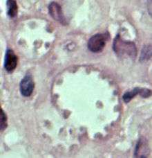 | |
| Leishmania mexicana in a biopsy specimen from a skin lesion stained with H&E. The amastigotes are lining the walls of two vacuoles, a typical arrangement. The species identification was derived from culture followed by isoenzyme analysis. | |
| Scientific classification | |
| Domain: | Eukaryota |
| (unranked): | Excavata |
| Phylum: | Euglenozoa |
| Class: | Kinetoplastida |
| Order: | Trypanosomatida |
| Genus: | Leishmania |
| Species: | L. mexicana |
| Binomial name | |
| Leishmania mexicana Biagi, 1953, emend. Garnham, 1962 | |

Leishmania mexicana belongs to Leishmania genus [1] and is the causal agent of cutaneous leishmaniasis in Mexico and central America .
Leishmania mexicana is an obligate intracellular protozoan parasite that causes the cutaneous form of leishmaniasis. This species of Leishmania is found in America. The infection with L. mexicana occurs when an individual is bitten by an infected sandfly that injects infective promastigotes, which are carried in the salivary glands and expulsed by the proboscis, directly to the skin.
The life cycle of this and other Leishmania species begin when an infected Phlebotomine bites and injects promastigotes in the skin of the mammal host. Those promastigotes are engulfed by phagocytic cells, as macrophages and dendritic cells. The parasites are kept inside in a parasitophorous vacuole, then they will transform to amastigotes and divide until brock the cell membrane. They are released to infect new cells as amastigotes stage. When an uninfected sand fly bites an infected mammal reservoir, the sandfly ingests the amastigotes, therefore they transform in promastigotes and divide in the midgut of the fly, those promastigotes migrates to the proboscis and are able to produce Leishmaniasis disease. There are no blood stages in the life cycle of L. mexicana (unlike Malaria and Trypanosomiasis).

L. mexicana can induce the cutaneous and diffuse cutaneous clinical manifestations in humans. The cutaneous type develops an ulcer at the bite site, here the amastigotes do not spread and the ulcers become visible either a few days or several months after the initial bite. The diffuse cutaneous type begins when the amastigote diffuses through the skin and metastasize to other tissue. This type does not produce ulcers and there is not treatment.
Treatment of Leishmaniasis caused by L. mexicana consists on pentavalent antimonials as Pentostam or Glucantime injected direct into the ulcer or Intramuscular.
Prevention of L. mexicana infection is principally avoiding the bitten of the sandfly.
References
- ↑ Majumdar D, Elsayed GA, Buskas T, Boons GJ (March 2005). "Synthesis of proteophosphoglycans of Leishmania major and Leishmania mexicana". J. Org. Chem. 70 (5): 1691–7. doi:10.1021/jo048443z. PMID 15730289.
- Vinetz JM, Soong L (February 2007). "Leishmania mexicana infection of the eyelid in a traveler to Belize". Braz J Infect Dis. 11 (1): 149–52. doi:10.1590/s1413-86702007000100030. PMID 17625744.
- Robertson CD, Coombs GH (February 1994). "Multiple high activity cysteine proteases of Leishmania mexicana are encoded by the Imcpb gene array". Microbiology (Reading, Engl.). 140 (Pt 2): 417–24. doi:10.1099/13500872-140-2-417. PMID 8180705.
- Ilg T, Etges R, Overath P, et al. (April 1992). "Structure of Leishmania mexicana lipophosphoglycan". J. Biol. Chem. 267 (10): 6834–40. PMID 1551890.
- Galindo-Sevilla N, Ortiz-Avalos J, Del Angel M, Galvan R, Mancilla-Ramirez J (2003). "Leishmania mexicana Strains Isolated from Both Localized Cutaneous (LCL) and Diffuse Cutaneous (DCL) Lesions in Humans Can Produce DCL in Mice, Being Faster in Males". ASM's Annual Meeting on Infectious Diseases: 43rd annual ICAAC Chicago 2003 : 43rd Interscience Conference on Antimicrobial Agents and Chemotherapy. American Society for Microbiology. ISBN 978-1-55581-284-3.
Pearson RD, Sousa AQ. Clinical spectrum of Leishmaniasis. Clin Infect Dis. 1996;22(1):1-13. PubMed PMID 8824958. Nat Rev Immunol. 2002 Nov;2(11):845-58. The immunology of susceptibility and resistance to Leishmania major in mice.