Calvaria (skull)
| Calvaria (skull) | |
|---|---|
 | |
| Details | |
| Identifiers | |
| Latin | calvaria |
| TA | A02.1.00.032 |
| FMA | 52800 |
The calvaria or skullcap (feminine Latin noun with plural calvariae; however, many medical texts list the word as calvarium, neuter Latin noun with plural calvaria) is the upper part of the neurocranium and covers the cranial cavity containing the brain.
The calvaria (skullcap) is made up of the superior portions of the frontal bone, occipital bone, and parietal bones.[1] In the human skull, the sutures between the bones normally remain flexible during the first few years of postnatal development, and fontanelles are palpable. Premature complete ossification of these sutures is called craniosynostosis.
Structure
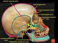
The outer surface of the skull possesses a number of landmarks. The point at which the frontal bone and the two parietal bones meet is known as "Bregma". The point at which the two parietal and occipital bones meet is known as "Lambda". Not only do these landmarks indicate the fontanelle in newborns, they also act as reference points in medicine and surgery.
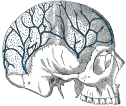
The inner surface of the skull-cap is concave and presents depressions for the convolutions of the cerebrum, together with numerous furrows for the lodgement of branches of the meningeal vessels. Along the middle line is a longitudinal groove, narrow in front, where it commences at the frontal crest, but broader behind; it lodges the superior sagittal sinus, and its margins afford attachment to the falx cerebri. On either side of it are several depressions for the arachnoid granulations, and at its back part, the openings of the parietal foramina when these are present.
It is crossed in front by the coronal suture and behind by the lambdoid suture, while the sagittal suture lies in the medial plane between the parietal bones.
Layers
Most bones of the calvaria consist of internal and external tables or layers of compact bone, separated by diploë. The diploë is cancellous bone containing red bone marrow during life, through which run canals formed by diploic veins. The diploë in a dried calvaria is not red because the protein was removed during preparation of the cranium. The internal table of bone is thinner than the external table, and in some areas there is only a thin plate of compact bone with no diploë.[2]


Development
In the fetus, the formation of the Calvaria involves a process known as intramembranous ossification.
References
- ↑ Tubbs, R. Shane; Bosmia, Anand N.; Cohen-Gadol, Aaron A. (27 November 2011). "The human calvaria: a review of embryology, anatomy, pathology, and molecular development". Child's Nervous System. 28 (1): 23–31. doi:10.1007/s00381-011-1637-0. PMID 22120469.
- ↑ Moore, K. L.; Daly, A. F.; Agur, A. M. R., eds. (2010). Clinically Oriented Anatomy (6th ed.). Baltimore: Lippincott Williams and Wilkins. p. 1168. ISBN 978-0-7817-7525-0.
Further reading
- Tubbs, R Shane; Loukas, Marios; Shoja, Mohammadali M; Apaydin, Nihal; Salter, E. George; Oakes, W. Jerry (April 2008) [20 Nov 2007]. "The intriguing history of the human calvaria: sinister and religious". Child's Nervous System. Germany: Springer-Verlag. 24 (4): 417–422. doi:10.1007/s00381-007-0509-0. ISSN 0256-7040. PMID 18026961.
External links
| Wikimedia Commons has media related to Calvaria (skull). |
- Cross section image: skull/calv-inf - Plastination Laboratory at the Medical University of Vienna
- Cross section image: skull/calv-sup - Plastination Laboratory at the Medical University of Vienna