Inferior longitudinal fasciculus
| Inferior longitudinal fasciculus | |
|---|---|
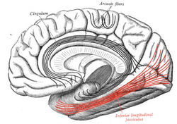 Medial surface of right cerebral hemisphere. Some of major association tracts are depicted. Inferior longitudinal fasciculus labeled at bottom right, in red. | |
 Diagram showing principal systems of association fibers in the cerebrum. (Inferior longitudinal fasciculus labeled at bottom right) | |
| Details | |
| Identifiers | |
| Latin | fasciculus longitudinalis inferior cerebri |
| NeuroNames | ancil-538 |
| TA | A14.1.09.556 |
| FMA | 77632 |
The inferior longitudinal fasciculus connects the temporal lobe and occipital lobe, running along the lateral walls of the inferior and posterior cornua of the lateral ventricle.
The existence of this fasciculus independent from the occipitotemporal fasciculus has been questioned for the human being, such that it has been proposed that the term inferior longitudinal fasciculus be replaced by the term "occipitotemporal projection".[1]
References
This article incorporates text in the public domain from the 20th edition of Gray's Anatomy (1918)
- ↑ Bergman, Ronald A.; Afifi, Adel K. (2005). Functional neuroanatomy: text and atlas. New York: McGraw-Hill. ISBN 0-07-140812-6.
External links
| Wikimedia Commons has media related to Inferior longitudinal fasciculus. |
- Atlas image: n1a5p6 at the University of Michigan Health System - "Dissection of the Left Hemisphere"
This article is issued from Wikipedia - version of the 6/1/2016. The text is available under the Creative Commons Attribution/Share Alike but additional terms may apply for the media files.