Immunoglobulin light chain
The immunoglobulin light chain is the small polypeptide subunit of an antibody (immunoglobulin).
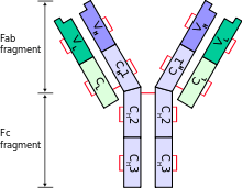
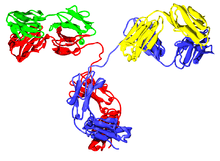
A typical antibody is composed of two immunoglobulin (Ig) heavy chains and two Ig light chains.
In humans
There are two types of light chain in humans (as in other mammals),
- kappa (κ) chain, encoded by the immunoglobulin kappa locus (IGK@) on chromosome 2
- lambda (λ) chain, encoded by the immunoglobulin lambda locus (IGL@) on chromosome 22
Antibodies are produced by B lymphocytes, each expressing only one class of light chain. Once set, light chain class remains fixed for the life of the B lymphocyte. In a healthy individual, the total kappa to lambda ratio is roughly 3:1 in serum (measuring intact whole antibodies) or 1:1.5 if measuring free light chains, with a highly divergent ratio indicative of neoplasm.
The exact normal ratio of kappa to lambda, according to a novel polyclonal free light chain assay, ranges from 0.26 to 1.65.[1] Both the kappa and the lambda chains can increase proportionately, maintaining a normal ratio. This is usually indicative of something other than a blood cell dyscrasia, such as kidney disease.
In other animals
The immunoglobulin light chain genes in tetrapods can be classified into three distinct groups: kappa (κ), lambda (λ), and sigma (σ). The divergence of the κ, λ, and σ isotypes preceded the radiation of tetrapods. The σ isotype was lost after the evolution of the amphibian lineage and before the emergence of the reptilian lineage.[2]
Other types of light chains can be found in lower vertebrates, such as the Ig-Light-Iota chain of Chondrichthyes and Teleostei.[3][4]
Camelids are unique among mammals as they also have fully functional antibodies which have two heavy chains, but lack the light chains usually paired with each heavy chain.[5] The functional role of this separate repertoire is unknown as yet.
Sharks also possess, as part of their adaptive immune systems, a functional heavy-chain homodimeric antibody-like molecule referred to as IgNAR (immunoglobulin new antigen receptor). IgNAR is believed to have never had an associated light chain, contrary to the understanding that the heavy-chain-only antibodies in camelids may have lost its light chain partner through evolution.[6]
Structure
Only one type of light chain is present in a typical antibody, thus the two light chains of an individual antibody are identical.
Each light chain is composed of two tandem immunoglobulin domains:
- one constant (CL) domain
- one variable domain (VL) that is important for binding antigen
The approximate length of a light chain protein is from 211 to 217 amino acids.[3] The constant region determines what class (kappa or lambda) the light chain is.[7] The lambda class has 4 subtypes (1, 2, 3, and 4).[7]

In pathology
Individual B-cells in lymphoid tissue possess either kappa or lambda light chains, but never both together. Specific rearrangement of lambda light chain of immunoglobulins can lead to loss of some protein coding genes, which does not seem to be functionally relevant (while functionally relevant miR-650 can be overexpressed).[8] Using immunohistochemistry, it is possible to determine the relative abundance of B-cells expressing kappa and lambda light chains. If the lymph node or similar tissue is reactive, or otherwise benign, it should possess a mixture of kappa positive and lambda positive cells. If, however, one type of light chain is significantly more common than the other, the cells are likely all derived from a small clonal population, which may indicate a malignant condition, such as B-cell lymphoma.[9]
Ig light chains produced in neoplastic plasma cells, such as in multiple myeloma, are called Bence Jones proteins.
Increased levels of free Ig light chains have also been detected in various inflammatory diseases. It is important to note that, in contrast to increased levels in lymphoma patients, these Ig light chains are polyclonal. Recent studies have shown that these Ig light chains can bind to mast cells and, using their ability to bind antigen, facilitate activation of these mast cells (Redegeld (2002)). Activation of mast cells results in the release of various pro-inflammatory mediators which are believed to contribute to the development of the inflammatory disease. Recent studies have shown that Ig light chains not only activate mast cells but also dorsal root ganglia (Rijnierse, 2009) and neutrophils (Braber and Thio (2012)), expanding their possible role as mediators in inflammatory disease.
See also
References
- ↑ Katzmann JA, Clark RJ, Abraham RS, Bryant S, Lymp JF, Bradwell AR, Kyle RA (2001). "Serum reference intervals and diagnostic ranges for free kappa and free lambda immunoglobulin light chains: relative sensitivity for detection of monoclonal light chains". Clin Chem. 48 (9): 1437–44. PMID 12194920.
- ↑ Das S, Nikolaidis N, Klein J, Nei M (2008). "Evolutionary redefinition of immunoglobulin light chain isotypes in tetrapods using molecular markers". Proc Natl Acad Sci U S A. 105 (43): 16647–52. doi:10.1073/pnas.0808800105. PMC 2575474
 . PMID 18940927.
 - 1 2 Janeway CA, Jr.; et al. (2001). Immunobiology. (5th ed.). Garland Publishing. (electronic full text via NCBI Bookshelf) ISBN 0-8153-3642-X.
- ↑ IMGT Index Antibodies (or Immunoglobulins).
- ↑ Hamers-Casterman C, Atarhouch T, Muyldermans S, Robinson G, Hamers C, Songa E, Bendahman N, Hamers R (1993). "Naturally occurring antibodies devoid of light chains". Nature. 363 (6428): 446–8. doi:10.1038/363446a0. PMID 8502296.
- ↑ Greenberg, A. S.; Avila, D.; Hughes, M.; Hughes, A.; McKinney, E. C.; Flajnik, M. F. (1995-03-09). "A new antigen receptor gene family that undergoes rearrangement and extensive somatic diversification in sharks". Nature. 374 (6518): 168–173. doi:10.1038/374168a0. ISSN 0028-0836. PMID 7877689.
- 1 2 Owen, Judith A.; Punt, Jenni; Stranford, Sharon (2013). Kuby Immunology. New York, NY: W. H. Freeman and Company. p. 85. ISBN 9781429219198.
- ↑ Mraz, M.; Stano Kozubik, K.; Plevova, K.; Musilova, K.; Tichy, B.; Borsky, M.; Kuglik, P.; Doubek, M.; Brychtova, Y.; Mayer, J.; Pospisilova, S. (2013). "The origin of deletion 22q11 in chronic lymphocytic leukemia is related to the rearrangement of immunoglobulin lambda light chain locus". Leukemia Research. 37 (7): 802–808. doi:10.1016/j.leukres.2013.03.018. PMID 23608880.
- ↑ Leong, Anthony S-Y; Cooper, Kumarason; Leong, F Joel W-M (2003). Manual of Diagnostic Cytology (2 ed.). Greenwich Medical Media, Ltd. pp. 283–285. ISBN 1-84110-100-1.
External links
- Immunoglobulin Light Chains at the US National Library of Medicine Medical Subject Headings (MeSH)
- Educational Resource for Immunoglobulin Light Chains