Hemodynamics
Hemodynamics or hæmodynamics is the dynamics of blood flow. The circulatory system is controlled by homeostatic mechanisms, much as hydraulic circuits are controlled by control systems. Hemodynamic response continuously monitors and adjusts to conditions in the body and its environment. Thus hemodynamics explains the physical laws that govern the flow of blood in the blood vessels. The relationships can be challenging because blood vessels are complex, with many ways for blood to enter and exit under changing conditions.
Cardiac output and flow rate

The heart is the driver of the circulatory system, pumping blood through rhythmic contraction and relaxation. The rate of blood flow out of the heart (often expressed in L/min) is known as the cardiac output (CO).
Blood being pumped out of the heart first enters the aorta, the largest artery of the body. It then proceeds to divide into smaller and smaller arteries, then into arterioles, and eventually capillaries, where oxygen transfer occurs. The capillaries connect to venules, and the blood then travels back through the network of veins to the right heart. The micro-circulation — the arterioles, capillaries, and venules —constitutes most of the area of the vascular system and is the site of the transfer of O2, glucose, and enzyme substrates into the cells. The venous system returns the de-oxygenated blood to the right heart where it is pumped into the lungs to become oxygenated and CO2 and other gaseous wastes exchanged and expelled during breathing. Blood then returns to the left side of the heart where it begins the process again.
In a normal circulatory system, the volume of blood returning to the heart each minute is approximately equal to the volume that is pumped out each minute (the cardiac output).[1] Because of this, the velocity of blood flow across each level of the circulatory system is primarily determined by the total cross-sectional area of that level. This is mathematically expressed by the following equation:
- v = Q/A
where
- v = velocity (cm/s)
- Q = blood flow (ml/s)
- A = cross sectional area (cm2)
Blood pressure across the circulatory system
The blood pressure in the circulation is principally due to the pumping action of the heart.[2] The pumping action of the heart generates pulsatile blood flow, which is conducted into the arteries, across the micro-circulation and eventually, back via the venous system to the heart. During each heartbeat, systemic arterial blood pressure varies between a maximum (systolic) and a minimum (diastolic) pressure.[3] In physiology these are often simplified into one value, the mean arterial pressure (MAP), which is calculated as follows:
- MAP ≈ 2⁄3(BPdia) + 1⁄3(BPsys)
where:
- MAP = Mean Arterial Pressure
- BPdia = Diastolic blood pressure
- BPsys = Systolic blood pressure
Differences in mean blood pressure are responsible for blood flow from one location to another in the circulation. The rate of mean blood flow depends on both blood pressure and the resistance to flow presented by the blood vessels. Mean blood pressure decreases as the circulating blood moves away from the heart through arteries and capillaries due to viscous losses of energy. Mean blood pressure drops over the whole circulation, although most of the fall occurs along the small arteries and arterioles.[4] Gravity affects blood pressure via hydrostatic forces (e.g., during standing), and valves in veins, breathing, and pumping from contraction of skeletal muscles also influence blood pressure in veins.[2]
The relationship between pressure, flow, and resistance is expressed in the following equation:[1]
- Flow = Pressure/Resistance
When applied to the circulatory system, we get:
- CO = (MAP – RAP)/TPR
where
- CO = cardiac output (in L/min)
- MAP = mean arterial pressure (in mmHg), the average pressure of blood as it leaves the heart
- RAP = right atrial pressure (in mmHg), the average pressure of blood as it returns to the heart
- TPR = total peripheral resistance (in mmHg * min/L)
A simplified form of this equation assumes right atrial pressure is approximately 0:
- CO ≈ MAP/TPR
The ideal blood pressure in the brachial artery, where standard blood pressure cuffs measure pressure, is <120/80 mmHg. Other major arteries have similar levels of blood pressure recordings indicating very low disparities among major arteries. In the innominate artery, the average reading is 110/70 mmHg, the right subclavian artery averages 120/80 and the abdominal aorta is 110/70 mmHg.[5] The relatively uniform pressure in the arteries indicate that these blood vessels act as a pressure reservoir for fluids that are transported within them.
Pressure drops gradually as blood flows from the major arteries, through the arterioles, the capillaries until blood is pushed up back into the heart via the venules, the veins through the vena cava with the help of the muscles. At any given pressure drop, the flow rate is determined by the resistance to the blood flow. In the arteries, with the absence of diseases, there is very little or no resistance to blood. The vessel diameter is the most principal determinant to control resistance. Compared to other smaller vessels in the body, the artery has a much bigger diameter (4 mm), therefore the resistance is low.[5]
The arm–leg (blood pressure) gradient is the difference between the blood pressure measured in the arms and that measured in the legs. It is normally less than 10 mm Hg,[6] but may be increased in e.g. coarctation of the aorta.[6]
Determinants of vascular resistance
Resistance is also related to vessel radius, vessel length, and blood viscosity.
In a first approach based on fluids, as indicated by the Hagen–Poiseuille equation.[7] The equation is as follows:
-
- ∆P: pressure drop/gradient
- µ: viscosity
- l: length of tube. In the case of vessels with infinitely long lengths, l is replaced with diameter of the vessel.
- Q: flow rate of the blood in the vessel
- r: radius of the vessel

In a second approach, more realistic of the vascular resistance and coming from experimental observations on blood flows, according to Thurston,[8] there is a plasma release-cell layering at the walls surrounding a plugged flow. It is a fluid layer in which at a distance δ, viscosity η is a function of δ written as η(δ), and these surrounding layers do not meet at the vessel centre in real blood flow. Instead, there is the plugged flow which is hyperviscous because holding high concentration of RBCs. Thurston assembled this layer to the flow resistance to describe blood flow by means of a viscosity η(δ) and thickness δ from the wall layer.
The blood resistance law appears as R adapted to blood flow profile :

where
- R = resistance to blood flow
- c = constant coefficient of flow
- L = length of the vessel
- η(δ) = viscosity of blood in the wall plasma release-cell layering
- r = radius of the blood vessel
- δ = distance in the plasma release-cell layer
Blood resistance varies depending on blood viscosity and its plugged flow (or sheath flow since they are complementary across the vessel section) size as well, and on the size of the vessels. Assuming steady, laminar flow in the vessel, the blood vessels behavior is similar to that of a pipe. For instance if p1 and p2 are pressures are at the ends of the tube, the pressure drop/gradient is:[9]

The larger arteries, including all large enough to see without magnification, are conduits with low vascular resistance (assuming no advanced atherosclerotic changes) with high flow rates that generate only small drops in pressure. The smaller arteries and arterioles have higher resistance, and confer the main blood pressure drop across major arteries to capillaries in the circulatory system.

In the arterioles blood pressure is lower than in the major arteries. This is due to bifurcations, which cause a drop in pressure. The more bifurcations, the higher the total cross-sectional area, therefore the pressure across the surface drops. This is why the arterioles have the highest pressure-drop. The pressure drop of the arterioles is the product of flow rate and resistance: ∆P=Q xresistance. The high resistance observed in the arterioles, which factor largely in the ∆P is a result of a smaller radius of about 30 µm.[10] The smaller the radius of a tube, the larger the resistance to fluid flow.
Immediately following the arterioles are the capillaries. Following the logic obvserved in the arterioles, we expect the blood pressure to be lower in the capillaries compared to the arterioles. Since pressure is a function of force per unit area, (P = F/A), the larger the surface area, the lesser the pressure when an external force acts on it. Though the radii of the capillaries are very small, the network of capillaries have the largest surface area in the vascular network. They are known to have the largest surface area (485 mm) in the human vascular network. The larger the total cross-sectional area, the lower the mean velocity as well as the pressure.[5]
Substances called vasoconstrictors can reduce the size of blood vessels, thereby increasing blood pressure. Vasodilators (such as nitroglycerin) increase the size of blood vessels, thereby decreasing arterial pressure.
If the blood viscosity increases (gets thicker), the result is an increase in arterial pressure. Certain medical conditions can change the viscosity of the blood. For instance, anemia (low red blood cell concentration), reduces viscosity, whereas increased red blood cell concentration increases viscosity. It had been thought that aspirin and related "blood thinner" drugs decreased the viscosity of blood, but instead studies found that they act by reducing the tendency of the blood to clot.[11]
Turbulence
Blood flow is also affected by the smoothness of the vessels, resulting in either turbulent (chaotic) or laminar (smooth) flow. Smoothness is reduced by the buildup of fatty deposits on the arterial walls.
The Reynold’s number (denoted NR or Re) is a relationship that helps determine the behavior of a fluid in a tube, in this case blood in the vessel.
The equation for this dimensionless relationship is written as:[7]
-
- ρ: density of the blood
- v: mean velocity of the blood
- L: characteristic dimension of the vessel, in this case diameter
- μ: viscosity of blood

The Reynold’s number is directly proportional to the velocity and diameter of the tube. Note that NR is directly proportional to the mean velocity as well as the diameter. A Reynold’s number of less than 2300 is laminar fluid flow, which is characterized by constant flow motion, whereas a value of over 4000, is represented as turbulent flow.[7] Due to its smaller radius and lowest velocity compared to other vessels, the Reynold’s number at the capillaries is very low, resulting in laminar instead of turbulent flow.[12]
Wall tension
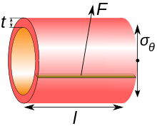
Regardless of site, blood pressure is related to the wall tension of the vessel according to the Young–Laplace equation (assuming that the thickness of the vessel wall is very small as compared to the diameter of the lumen):

where
- P is the blood pressure
- t is the wall thickness
- r is the inside radius of the cylinder.
- is the cylinder stress or "hoop stress".

For the thin-walled assumption to be valid the vessel must have a wall thickness of no more than about one-tenth (often cited as one twentieth) of its radius.
The cylinder stress, in turn, is the average force exerted circumferentially (perpendicular both to the axis and to the radius of the object) in the cylinder wall, and can be described as:

where:
- F is the force exerted circumferentially on an area of the cylinder wall that has the following two lengths as sides:
- t is the radial thickness of the cylinder
- l is the axial length of the cylinder
Venous capacitance
Veins are described as the "capacitance vessels" of the body because over 70% of the blood volume resides in the venous system. Veins are more compliant than arteries and expand to accommodate changing volume.[13]
Monitoring
Hemodynamic monitoring is the observation of hemodynamic parameters over time, such as blood pressure and heart rate. Blood pressure can be monitored either invasively through an inserted blood pressure transducer assembly (providing continuous monitoring), or noninvasively by repeatedly measuring the blood pressure with an inflatable blood pressure cuff.
Etymology and pronunciation
The word hemodynamics (/ˌhiːmədaɪˈnæmɪks, -moʊ-/[14]) uses combining forms of hemo- and dynamics, thus "the dynamics of blood". The vowel of the hemo- syllable is variously written according to the ae/e variation.
See also
- Cardiac output
- Blood pressure
- Blood flow
- Electrical cardiometry
- Photoplethysmograph
- Impedance cardiography
- Esophogeal doppler
- Blood hammer
- Windkessel effect
Notes and references
- 1 2 Costanzo, Linda S. (2003). Physiology. Board Review Series (3rd ed.). Philadelphia: Lippincott Williams and Wilkins. pp. 73–113. ISBN 0781739195.
- 1 2 Caro, Colin G. (1978). The Mechanics of The Circulation. Oxford [Oxfordshire]: Oxford University Press. ISBN 0-19-263323-6.
- ↑ "Normal Blood Pressure Range Adults". Health and Life.
- ↑ Klabunde, Richard (2005). Cardiovascular Physiology Concepts. Lippincott Williams & Wilkins. pp. 93–4. ISBN 978-0-7817-5030-1.
- 1 2 3 Fung, Yuan-cheng (1997). Biomechanics:Circulation. New York: Springer. p. 571. ISBN 0-387-94384-6.
- 1 2 Markham LW, Knecht SK, Daniels SR, Mays WA, Khoury PR, Knilans TK (November 2004). "Development of exercise-induced arm-leg blood pressure gradient and abnormal arterial compliance in patients with repaired coarctation of the aorta". Am. J. Cardiol. 94 (9): 1200–2. doi:10.1016/j.amjcard.2004.07.097. PMID 15518624.
- 1 2 3 Munson BR, Young DF, Okiishi TH, Huebsch WW (2009). Fundamentals of Fluid Mechanics (Sixth ed.). New Jersey: John Wiley &Sons, Inc. p. 725. ISBN 978-0-470-26284-9.
- 1 2 GB Thurston, Viscosity and viscoelasticity of blood in small diameter tubes, Microvasular Research 11, 133 146, 1976
- ↑ Womersley JR (1955). "Method for the calculation of velocity, rate of flow and viscous drag in arteries when the pressure gradient is known". Journal of Physiology. 127 (3): 553–63. PMC 1365740
 . PMID 14368548.
 - ↑ Sircar, Sabyasach (2008). Principles of Medical Physiology. India: vistasta Publishing. ISBN 978-1-58890-572-7.
- ↑ Rosenson RS, Wolff D, Green D, Boss AH, Kensey KR (February 2004). "Aspirin. Aspirin does not alter native blood viscosity". J. Thromb. Haemost. 2 (2): 340–1. doi:10.1111/j.1538-79333.2004.0615f.x. PMID 14996003.
- ↑ Fung, Yuan-cheng; Zweifach, B.W. (1971). "Microcirculation: Mechanics of Blood Flow in Capillaries". Annual Review of Fluid Mechanics. 3: 189–210. Bibcode:1971AnRFM...3..189F. doi:10.1146/annurev.fl.03.010171.001201.
- ↑ Lough, Mary (2015-04-15). Hemodynamic Monitoring: Evolving technologies and clinical practice (1 ed.). St. Louis, Missouri: Elsevier Mosby. p. 25. ISBN 978-0-323-08512-0.
- ↑ "haemodynamic". Oxford Dictionaries. Oxford University Press. Retrieved 2016-01-20.
Bibliography
- Berne RM, Levy MN. Cardiovascular physiology. 7th Ed Mosby 1997
- Rowell LB. Human Cardiovascular Control. Oxford University press 1993
- Braunwald E (Editor). Heart Disease: A Textbook of Cardiovascular Medicine. 5th Ed. W.B.Saunders 1997
- Siderman S, Beyar R, Kleber AG. Cardiac Electrophysiology, Circulation and Transport. Kluwer Academic Publishers 1991
- American Heart Association
- Otto CM, Stoddard M, Waggoner A, Zoghbi WA. Recommendations for Quantification of Doppler Echocardiography: A Report from the Doppler Quantification Task Force of the Nomenclature and Standards Committee of the American Society of Echocardiography. J Am Soc Echocardiogr 2002;15:167-184
- Peterson LH, The Dynamics of Pulsatile Blood Flow, Circ. Res. 1954;2;127-139
- Hemodynamic Monitoring, Bigatello LM, George E., Minerva Anestesiol, 2002 Apr;68(4):219-25
- Claude Franceschi; Paolo Zamboni Principles of Venous Hemodynamics Nova Science Publishers 2009-01 ISBN Nr 1606924850/9781606924853
- WR Milnor: Hemodynamics, Williams & Wilkins, 1982
- B Bo Sramek: Systemic Hemodynamics and Hemodynamic Management, 4th Edition, ESBN 1-59196-046-0
External links
- Hemodynamic Society
- Learn hemodynamics
- Educational Particle Image Velocimetry (e-PIV) - resources and demonstrations
| Library resources about Hemodynamics |