Gallbladder
| Gallbladder | |
|---|---|
.png) | |
| Details | |
| Precursor | Foregut |
| System | Digestive system |
| Artery | Cystic artery |
| Vein | Cystic vein |
| Nerve | Celiac ganglia, Vagus (CN X)[1] |
| Identifiers | |
| Latin | Vesica biliaris, vesica fellea |
| MeSH | A03.159.439 |
| TA | A05.8.02.001 |
| FMA | 7202 |
In vertebrates the gallbladder (also gall bladder, biliary vesicle or cholecyst) is a small organ where bile (a fluid produced by the liver) is stored and concentrated before it is released into the small intestine. Humans can live without a gallbladder. The surgical removal of the gallbladder is called a cholecystectomy.
Structure

The gallbladder is a hollow organ that sits just beneath the right lobe of the liver.[2] In adults, the gallbladder measures approximately 8 centimetres (3.1 inches) in length and 4 centimetres (1.6 in) in diameter when fully distended.[3] The gallbladder has a capacity of about 100 millilitres (3.5 imperial fluid ounces).[4]:298
The gallbladder is shaped like a tapered sac, with the open end opening into the biliary tree and the cystic duct. Anatomically, the gallbladder is divided into three sections: the fundus, body, and neck:[5] The fundus is a rounded end that faces the front of the body.[5] The body is in contact with the liver, lying in the gallbladder fossa, a depression at the bottom of the liver.[5] The neck tapers and is continuous with the cystic duct, part of the biliary tree. The gallbladder fossa, against which the fundus and body of the gallbladder lie, is found beneath the junction of hepatic segments IVB and V.[6] The cystic duct unites with the common hepatic duct to become the common bile duct. At the junction of the neck of the gallbladder and the cystic duct, there is an out-pouching of the gallbladder wall forming a mucosal fold known as Hartmann's pouch, where gallstones commonly get stuck.
The angle of the gallbladder is located between the costal margin and the lateral margin of the rectus abdominis muscle. The fundus is at the same level as the transpyloric plane.
Histology

The layers of the gallbladder wall are visible under the microscope. The gallbladder wall's innermost surface is lined by a single layer of columnar cells with an apical brush border of microvilli, very similar to intestinal absorptive cells.[7][8] Underneath the epithelia is an underlying lamina propria, a muscular layer, an outer perimuscular layer and serosa. Unlike elsewhere in the intestinal tract, the gallbladder does not have a muscularis mucosae, and the muscular fibres are not arranged in distinct layers.[4] In greater detail, the layers are:[4][9]
- The epithelium is the innermost layer of the gallbladder, and is of simple columnar type. Underneath the epithelium is a lamina propria; together, these two layers are known as the mucosa. A distinctive feature of the gallbladder is the presence of Rokitansky–Aschoff sinuses, deep outpouchings of the mucosa that can extend through the muscular layer.[10]
- The submucosa is a thin layer of loose connective tissue with smaller blood vessels. It contains many elastin fibres, lymphatics, and in the neck of the gallbladder, glands which secrete mucus. The lymphatics of this layer help to drain water when the bile is concentrated, and the mucous glands may create a surface that protects the wall of the biliary tree.
- The muscular layer, formed by smooth muscular tissue. The interspersed muscle fibres lie in longitudinal, oblique and transverse directions, and are not arranged in separate layers. The muscle fibres here contract to expel bile from the gallbladder.
- The perimuscular ("around the muscle") fibrous tissue, another layer of connective tissue
- The serosa is a thick layer that covers the outer surface of the gallbladder, and is continuous with the peritoneum, which lines the abdominal cavity. The serosa contains blood vessels and lymphatics.
Development
The gallbladder develops from an endodermal outpouching of the embryonic gut tube.[11] Early in development, the human embryo has three germ layers and abuts an embryonic yolk sac. During the second week of embryogenesis, as the embryo grows, it begins to surround and envelop portions of this sac. The enveloped portions form the basis for the adult gastrointestinal tract. Sections of this foregut begin to differentiate into the organs of the gastrointestinal tract, such as the oesophagus, stomach, and intestines.[11]
During the fourth week of embryological development, the stomach rotates. The stomach, originally lying in the midline of the embryo, rotates so that its body is on the left. This rotation also affects the part of the gastrointestinal tube immediately below the stomach, which will go on to become the duodenum. By the end of the fourth week, the developing duodenum begins to spout a small outpouching on its right side, the hepatic diverticulum, which will go on to become the biliary tree. Just below this is a second outpouching, known as the cystic diverticulum, that will eventually develop into the gallbladder.[11]
Variation
Anatomical variants of the gallbladder occur very rarely, although a range of abnormalities have been documented.
The number and structure of the gallbladder may vary. Occasionally two or even three gallbladders may coexist, either as separate bladders draining into the cystic duct, or sharing a common branch that drains into the cystic duct. Additionally, the gallbladder may fail to form at all. Gallbladders with two lobes separated by a septum may also exist. These abnormalities are not likely to affect function and are generally asymptomatic.[12]
The location of the gallbladder in relation to the liver may also vary, with documented variants including gallbladders found within,[13] above, on the left side of, behind, and detached from the liver. Such variants are very rare: from 1886 to 1998, only 110 cases of left-lying liver, or less than one per year, were reported in scientific literature.[14][15]
An anatomical variation can occur, known as a Phrygian cap, which is an innocuous fold in the fundus, named after its resemblance to the Phrygian cap.[16]
Function
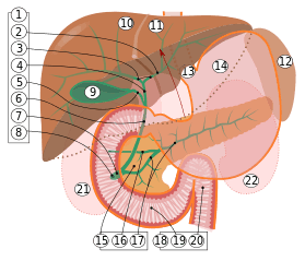
9. Gallbladder, 10–11. Right and left lobes of liver. 12. Spleen.
13. Esophagus. 14. Stomach. 15. Pancreas: 16: Accessory pancreatic duct, 17: Pancreatic duct.
18. Small intestine: 19. Duodenum, 20. Jejunum
21–22: Right and left kidneys (silhouette).
The anterior border of the liver is lifted upwards (brown arrow). Gallbladder with Longitudinal section, pancreas and duodenum with frontal one. Intrahepatic ducts and stomach in transparency.
The main purpose of the gallbladder is to store bile, also called gall, needed for the digestion of food. The gallbladder is part of the biliary system and serves as a reservoir for bile, which is produced by the liver. The liver produces the bile, which then flows through the hepatic ducts into the gallbladder. At any one time, 30 to 60 millilitres (1.0 to 2.0 US fl oz) of bile is stored within the gallbladder.[17]
When food containing fat enters the digestive tract, it stimulates the secretion of cholecystokinin (CCK) from I cells of the duodenum and jejunum. In response to cholecystokinin, the gallbladder rhythmically contracts and releases its contents into the common bile duct, eventually draining into the duodenum. The bile emulsifies fats in partly digested food, thereby assisting their absorption. Bile consists primarily of water and bile salts, and also acts as a means of eliminating bilirubin, a product of hemoglobin metabolism, from the body.[17]
The bile that is secreted by the liver and stored in the gallbladder is not the same as the bile that is secreted by the gallbladder. During gallbladder storage of bile, it is concentrated by removal of some water and electrolytes. This is through the active transport of sodium ions across the epithelia of the gallbladder, which creates an osmotic pressure that also causes water and other electrolytes such as chloride to be reabsorbed.[17]
Clinical significance
Gallstones
Gallstones are the most common problem to affect the gallbladder.[18] Gallstones generally form when the bile is saturated with either cholesterol or bilirubin. Only a minority of gallstones cause symptoms; most stones are passed along the biliary system. When symptoms occur, severe pain in the upper right part of the abdomen is felt. If the stone blocks the gallbladder, inflammation as cholecystitis may result. If the stone lodges in the biliary system, jaundice may occur; and if the stone blocks the pancreatic duct, then pancreatitis may occur. Gallstones are often managed by waiting for them to be naturally passed. In people with recurrent gallstones, surgery to remove the gallbladder may be considered. Some medication, such as ursodeoxycholic acid, may be used; and lithotripsy, a procedure used to break down the stones, may also be used.[19]
Inflammation
Inflammation of the gallbladder is known as cholecystitis. Inflammation is most commonly because of obstruction of the duct with gallstones, known as cholelithiasis. Blocked bile accumulates, and pressure on the gallbladder wall may lead to the release of substances that cause inflammation, such as phospholipase. There is also the risk of bacterial infection. An inflamed gallbladder is likely to cause pain and fever, and tenderness in the upper, right corner of the abdomen, and may have a positive Murphy's sign. Cholecystitis is often managed with rest and antibiotics, particularly cephalosporins and, in severe cases, metronidazole.[19]
Cholecystitis may also occur chronically, particularly when a person is prone to getting gallstones.[19]
Cholesterolosis
Cholesterolosis of the gallbladder, also called strawberry gallbladder, is a change in the gallbladder wall due to excess cholesterol.[20] It is not linked to gallstones or inflammation.
Gallbladder polyps
Gallbladder polyps are mostly benign growths or lesions resembling growths that form in the gallbladder wall.[21]
Gallbladder removal
A cholecystectomy is a procedure in which the gallbladder is removed. It may be removed because of recurrent gallstones, and is considered an elective procedure. A cholecystectomy may be an open procedure, or one conducted by laparoscopy. In the surgery, the gallbladder is removed from the neck to the fundus,[22] and so bile will drain directly from the liver into the biliary tree. About 30 percent of patients may experience some degree of indigestion following the procedure, although severe complications are much rarer.[19]
About 10 percent of surgeries lead to a chronic condition of postcholecystectomy syndrome.[23][24]
Imaging

Ultrasound is often the first imaging examination performed when galbladder disease is suspected. Other imaging options include MRCP (magnetic resonance cholangiopancreatography), ERCP and percutaneous or intraoperative cholangiography. A cholescintigraphy scan is a nuclear imaging procedure used to assess the condition of the gallbladder.
Society and culture
Numerous words in the English language relate to the gallbladder and the bile that it stores. To have 'gall' is associated with bold behaviour, whereas to have 'bile' is associated with bitterness.[25]
In the Chinese language, the gallbladder (Chinese: 膽) is associated with courage and a plethora of related idioms, including using terms such as "a body completely [of] gall" (Chinese: 渾身是膽) to describe a brave person, and "single gallbladder hero" (Chinese: 孤膽英雄) to describe a lone hero.[26]
In the Zangfu theory of Chinese medicine, the gallbladder not only has a digestive role, but is seen as the seat of decision-making.[26]
Bile is commonly misinterpreted to be stomach acid; rather, it is secreted into the duodenum.
Other animals
Most vertebrates have gallbladders, but the form and arrangement of the bile ducts may vary considerably. In many species, for example, there are several separate ducts running to the intestine, rather than the single common bile duct found in humans. Several species of mammals (including horses, deer, rats, and laminoids),[27][28] several species of birds, lampreys and all invertebrates lack a gallbladder altogether.[29]
Several species of bears are farmed or hunted for their gallbladders or bile.
See also
References
- ↑ Ginsburg, Ph.D., J.N. (August 22, 2005). "Control of Gastrointestinal Function". In Thomas M. Nosek, Ph.D. Gastrointestinal Physiology. Essentials of Human Physiology. Augusta, Georgia, United State: Medical College of Georgia. pp. p. 30. Retrieved June 29, 2007.
- ↑ "Where is the Gallbladder Located in the Body". Buzzle.com. February 28, 2013. Retrieved August 18, 2013.
- ↑ Jon W. Meilstrup (1994). Imaging Atlas of the Normal Gallbladder and Its Variants. Boca Raton: CRC Press. p. 4. ISBN 0-8493-4788-2.
- 1 2 3 Young, Barbara; et al. (2006). Wheater's functional histology: a text and colour atlas (5th ed.). [Edinburgh?]: Churchill Livingstone/Elsevier. p. 298. ISBN 978-0-443-06850-8.
- 1 2 3 Drake, Richard L.; Vogl, Wayne; Tibbitts, Adam; Mitchell, W.M.; Richard (illustrations); Richardson, Paul (2005). Gray's anatomy for students. Philadelphia: Elsevier/Churchill Livingstone. p. 287. ISBN 978-0-8089-2306-0.
- ↑ Shakelford's Surgery of Alimentary Tract, ed.7. 2013
- ↑ "Gall bladder". Retrieved April 15, 2014.
- ↑ Zaki, Mohamed; Al-Refeidi, Abdullah (2009). "Histological Changes in the Human Gallbladder Epithelium associated with Gallstones". OMJ. 24: 269–273. doi:10.5001/omj.2009.55.
- ↑ "Staging of Gallbladder Cancer".
- ↑ Ross, M.; Pawlina, W. (2011). Histology: A Text and Atlas (6th ed.). Lippincott Williams & Wilkins. p. 646. ISBN 978-0-7817-7200-6.
- 1 2 3 Gary C. Schoenwolf; et al. (2009). Larsen's human embryology (Thoroughly rev. and updated 4th ed.). Philadelphia: Churchill Livingstone/Elsevier. pp. "Development of the Gastrointestinal Tract". ISBN 978-0-443-06811-9.
- ↑ Leeuw, Th.G.; Verbeek, P.C.M.; Rauws, E.A.J.; Gouma, D.J. (September 1995). "A double or bilobar gallbladder as a cause of severe complications after (laparoscopic) cholecystectomy". Surgical Endoscopy. 9 (9): 998–1000. doi:10.1007/BF00188459. PMID 7482221.
- ↑ Segura-Sampedro, JJ; Navarro-Sánchez, A; Ashrafian, H; Martínez-Isla, A (February 2015). "Laparoscopic approach to the intrahepatic gallbladder. A case report.". Revista espanola de enfermedades digestivas : organo oficial de la Sociedad Espanola de Patologia Digestiva. 107 (2): 122–3. PMID 25659400.
- ↑ Dhulkotia, A; Kumar, S; Kabra, V; Shukla, HS (March 1, 2002). "Aberrant gallbladder situated beneath the left lobe of liver". HPB. 4 (1): 39–42. doi:10.1080/136518202753598726.
- ↑ Naganuma, S.; Ishida, H.; Konno, K.; Hamashima, Y.; Hoshino, T.; Naganuma, H.; Komatsuda, T.; Ohyama, Y.; Yamada, N.; Ishida, J.; Masamune, O. (March 6, 2014). "Sonographic findings of anomalous position of the gallbladder". Abdominal Imaging. 23 (1): 67–72. doi:10.1007/s002619900287. PMID 9437066.
- ↑ Meilstrup JW; Hopper KD; Thieme GA (December 1991). "Imaging of gallbladder variants". AJR Am J Roentgenol. 157 (6): 1205–8. doi:10.2214/ajr.157.6.1950867. PMID 1950867.
- 1 2 3 Hall, Arthur C. Guyton, John E. (2005). Textbook of medical physiology (11th ed.). Philadelphia: W.B. Saunders. pp. 802–804. ISBN 978-0-7216-0240-0.
- ↑ Rodriguez, D. (January 25, 2010). What Is the Gallbladder?. Everyday Health, Retrieved March 20, 2011, from http://www.everydayhealth.com/gallbladder/what-is-the-gallbladder.html
- 1 2 3 4 Britton, the editors Nicki R. Colledge, Brian R. Walker, Stuart H. Ralston ; illustrated by Robert (2010). Davidson's principles and practice of medicine. (21st ed.). Edinburgh: Churchill Livingstone/Elsevier. pp. 977–979. ISBN 978-0-7020-3085-7.
- ↑ Strawberry gallbladder – cancerweb.ncl.ac.uk.
- ↑ "Gallbladder Polyps". MayoClinic. Retrieved March 19, 2015.
- ↑ Neri V; Ambrosi A; Fersini A; Tartaglia N; Valentino TP (2007). "Antegrade dissection in laparoscopic cholecystectomy". JSLS: Journal of the Society of Laparoendoscopic Surgeons / Society of Laparoendoscopic Surgeons. 11 (2): 225–8. PMC 3015719
 . PMID 17761085.
 - ↑ Surcigal Science, Post Cholecystectomy Diarrhoea—A Systematic Review, 2012
- ↑ nhs.uk, Complications of a gallbladder removal
- ↑ J. A. Simpson (1989). The Oxford English dictionary (2nd ed.). Oxford: Clarendon Press. gall, bile. ISBN 978-0-19-861186-8.
- 1 2 Yu, Ning (January 1, 2003). "Metaphor, Body, and Culture: The Chinese Understanding of Gallbladder and Courage". Metaphor and Symbol. 18 (1): 13–31. doi:10.1207/S15327868MS1801_2.
- ↑ C. Michael Hogan. 2008. Guanaco: Lama guanicoe, GlobalTwitcher.com, ed. N. Strömberg
- ↑ Higashiyama H, Sumitomo H, Ozawa A, Igarashi H, Tsunekawa N, Kurohmaru M, Kanai Y. (2016). Anatomy of the Murine Hepatobiliary System: A Whole-Organ-Level Analysis Using a Transparency Method. The Anatomical Record. 299(2):161-172. doi:10.1002/ar.23287 PMID 26559382
- ↑ Romer, Alfred Sherwood; Parsons, Thomas S. (1977). The Vertebrate Body. Philadelphia, PA: Holt-Saunders International. p. 355. ISBN 0-03-910284-X.
External links
| Wikimedia Commons has media related to Gallbladder. |
- Diagram of Human Stomach and Gallbladder – Human Anatomy Online dd, MyHealthScore.com.
- www.newchronicles.webs.com/f/gastrointestinalphysiology – Gastrointestinal Physiology Review.
- http://www.ece.ncsu.edu/imaging/MedImg/SIMS/GF32.gif
- Anatomy photo:38:14-0100 at the SUNY Downstate Medical Center - "Stomach, Spleen and Liver: The Gallbladder and the Bile System"
- Rodriguez, D. (January 25, 2010). "What Is the Gallbladder?" Everyday Health. Retrieved July 9, 2015.
- "Life Without a Gallbladder". Digestive Disorders (January 2009), 30–31. Retrieved n.d., from Health Source – Consumer Edition (ISBN 978-0-929661-67-4).