Cardiac skeleton
The cardiac skeleton, also known as the fibrous skeleton of the heart, is a high density single structure of connective tissue that forms and anchors the valves and influences the forces exerted through them. The cardiac skeleton separates and partitions the atria (the smaller, upper two chambers) from the ventricles (the larger, lower two chambers). This is important because it forms the primary channel that electrical energy follows from the top to the bottom of the heart.
Structure
The cardiac skeleton consists of four bands of dense connective tissue, as collagen, that encircle the bases of the pulmonary trunk, aorta, and heart valves.[1] While not a "true" skeleton, it does provide structure and support for the heart, as well as isolating the atria from the ventricles. In youth, this collagen structure is free of calcium adhesions and is quite flexible. With aging some calcium can accumulate on this skeleton. This accumulation contributes to the delay of the depolarisation wave in geriatric patients that can take place from the AV node and the bundle of His.[2]
Fibrous rings
| Fibrous rings of heart | |
|---|---|
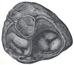 Transverse section of the heart showing the fibrous rings surrounding the valves | |
| Details | |
| Latin | anulus fibrosus dexter cordis, anulus fibrosus sinister cordis |
| Fibrous trigone | |
|---|---|
| Details | |
| Latin | trigonum fibrosum dextrum cordis, trigonum fibrosum sinistrum cordis, trigona fibrosa |
The right and left fibrous rings of heart (anulus fibrosus cordis) surround the atrioventricular and arterial orifices. The right fibrous ring is known as the anulus fibrosus dexter cordis, and the left is known as the anulus fibrosus sinister cordis.[2] The right fibrous trigone is continuous with the central fibrous body. This is the strongest part of the fibrous cardiac skeleton.
The upper chambers (atria) and lower (ventricles) are electrically divided by the properties of collagen proteins within the rings. The valve rings, central body and skeleton of the heart consisting of collagen are impermeable to electrical propagation. The only channel allowed (barring accessory/rare preexcitation channels) through this collagen barrier is represented by a sinus that opens up to the atrioventricular node and exits to the bundle of His. The muscle origins/insertions of many of the cardiomyocytes are anchored to opposite sides of the valve rings.[2]
The atrioventricular rings serve for the attachment of the muscular fibers of the atria and ventricles, and for the attachment of the bicuspid and tricuspid valves.[2]
The left atrioventricular ring is closely connected, by its right margin, with the aortic arterial ring; between these and the right atrioventricular ring is a triangular mass of fibrous tissue, the Fibrous trigone, which represents the os cordis seen in the heart of some of the larger animals, as the ox and elephant.[2]
Lastly, there is the tendinous band, already referred to, the posterior surface of the conus arteriosus.[2]
The fibrous rings surrounding the arterial orifices serve for the attachment of the great vessels and semilunar valves, they are known as The aortic annulus.[2]
Each ring receives, by its ventricular margin, the attachment of some of the muscular fibers of the ventricles; its opposite margin presents three deep semicircular notches, to which the middle coat of the artery is firmly fixed.[2]
The attachment of the artery to its fibrous ring is strengthened by the external coat and serous membrane externally, and by the endocardium internally.[2]
From the margins of the semicircular notches the fibrous structure of the ring is continued into the segments of the valves.[2]
The middle coat of the artery in this situation is thin, and the vessel is dilated to form the sinuses of the aorta and pulmonary artery.[2]
Function
Electrical signals from the sinoatrial node and the autonomic nervous system must find their way from the upper chambers to the lower ones to ensure that the ventricles can drive the flow of blood. The heart functions as a pump delivering an intermittent volume of blood, incrementally delivered to the lungs, body and brain.
The cardiac skeleton ensures that the electrical and autonomic energy generated above is ushered below and cannot return. The cardiac skeleton does this by establishing an electrically impermeable boundary to autonomic electrical influence within the heart. Simply put, the dense connective tissue within the cardiac skeleton does not conduct electricity and its deposition within the myocardial matrix is not accidental.
The anchored and electrically inert collagen framework of the four valves allows normal anatomy to house the atrioventricular node (AV node) in its center. The AV node is the only electrical conduit from the atria to the ventricles through the cardiac skeleton, which is why atrial fibrillation can never degrade into ventricular fibrillation.
Throughout life, the cardiac collagen skeleton is remodeled. Where collagen is diminished by age, calcium is often deposited, thus allowing readily imaged mathematical markers which are especially valuable in measuring systolic volumetrics. The inert characteristics of the collagen structure that blocks electrical influence also makes it difficult to attain an accurate signal for imaging without allowing for an applied ratio of collagen to calcium.
History
Boundaries within the heart were first described and greatly magnified by Drs. Charles S. Peskin and David M. McQueen at the Courant Institute of Mathematical Sciences.
See also
References
This article incorporates text in the public domain from the 20th edition of Gray's Anatomy (1918)
External links
- -1026555847 at GPnotebook
- Description at cwc.net
- 261750843 at GPnotebook, – "left fibrous trigone"
- 798621756 at GPnotebook, – "right fibrous trigone"
- Histology (see slide #96)