Hip dysplasia
| Hip dysplasia (human) | |
|---|---|
 | |
| Congenital dislocation of the left hip in an elderly person. Closed arrow marks the acetabulum, open arrow the femoral head. | |
| Classification and external resources | |
| ICD-10 | Q65 |
| ICD-9-CM | 754.3 |
| OMIM | 142700 |
| DiseasesDB | 3056 |
| MedlinePlus | 000971 |
| eMedicine | orthoped/456 |
| MeSH | D006618 |
Hip dysplasia, developmental dysplasia of the hip (DDH)[1] or congenital dysplasia of the hip (CDH)[2] is a congenital or developmental deformation or misalignment of the hip joint.
Signs and symptoms
Hip dysplasia can range from barely detectable to severely malformed or dislocated. The congenital form, teratologic or non-reducible dislocation occurs as part of more complex conditions.
The condition can be bilateral or unilateral:
- If both hip joints are affected one speaks of "bilateral" dysplasia. In this case some diagnostic indicators like asymmetric folds and leg-length inequality do not apply.
- In unilateral dysplasia only one joint shows deformity, the contralateral side may show resulting effects.[3] In the majority of unilateral cases the left hip has the dysplasia.
If the joint is fully dislocated a false acetabulum often forms (often higher up on the pelvis) opposite the dislocated femoral head position.
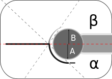
In acetabular dysplasia the acetabulum (socket) is too shallow or deformed. The center-edge angle is measured as described by Wiberg.[4] Two forms of femoral dysplasia are coxa vara, in which the femur head grows at too narrow an angle to the shaft, and coxa valga, in which the angle is too wide.
A rare type, the "Beukes familial hip dysplasia" is found among Afrikaners that are members of the Beukes family. The femur head is flat and irregular. People develop osteoarthritis at an early age.[5]
Causes
Hip dysplasia is considered to be a multifactorial condition. That means that several factors are involved in causing the condition to manifest.[6] Its cause is unknown but it is common in cases of a large fetus or a fetus in a breech position.
Congenital
Some studies suggest a hormonal link.[7] Specifically, the hormone relaxin has been indicated.[8]
A genetic factor is indicated since the trait runs in families and there is an increased occurrence in some ethnic populations (e.g., Native Americans,[9] Lapps[10] / Sami people[11]). A locus has been described on chromosome 13.[12] Beukes familial dysplasia, on the other hand, was found to map to an 11-cM region on chromosome 4q35, with nonpenetrant carriers not affected.[13]
Acquired
As an acquired condition it has often been linked to traditions of swaddling infants,[14] use of overly restrictive baby seats, carriers and other methods of transporting babies,[15] or use of a cradle board which locks the hip joint in an "adducted" position (pulling the knees together tends to pull the heads of the femur bone out of the sockets or acetabulae) for extended periods. Modern swaddling techniques, such as the 'hip healthy swaddle' have been developed to relieve stress on hip joints caused by traditional swaddling methods.[16]
Further risk factors include breech birth, gender, genetics (family history),[17] and firstborns.[18] In breech position the femoral head tends to get pushed out of the socket. A narrow uterus also facilitates hip joint dislocation during fetal development and birth.
Diagnosis
Most countries have standard newborn exams that include a hip joint exam screening for early detection of hip dysplasia. Sometimes during an exam a "click" or more precisely "clunk" in the hip may be detected[19] (although not all clicks indicate hip dysplasia).[20] When a hip click (also known as "clicky hips" in the UK) is detected, the child's hips are tracked with additional screenings[21] to determine if developmental dysplasia of the hip is caused.[22]
Two maneuvers commonly employed for diagnosis in neonatal exams are the Ortolani maneuver and the Barlow maneuver.[23]
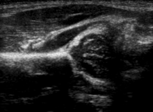
The condition can be confirmed by ultrasound[24] and X-ray.[25] Ultrasound imaging yields better results defining the anatomy until the cartilage is ossified. When the infant is around 3 months old a clear roentgenographic image can be achieved. Unfortunately the time the joint gives a good x-ray image is also the point at which nonsurgical treatment methods cease to give good results. In x-ray imaging dislocation may be indicated if the Shenton's line (an arc drawn from the medial aspect of the femoral neck through the superior margin of the obturator foramen[26]) does not result in a smooth arc. However, in infants this line can be unreliable as it depends on the rotation of the hip when the image is taken ([27])
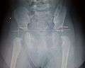 X-Ray showing calculations for working out hip dysplasia
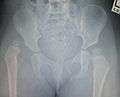 X-Ray Image showing Hip Dysplasia in an Infant
Asymmetrical gluteal folds and an apparent limb-length inequality can further indicate unilateral hip dysplasia.[28] Most vexingly, many newborn hips show a certain ligamentous laxity, on the other hand severely malformed joints can appear stable. That is one reason why follow-up exams and developmental monitoring are important. Frequency and methods of routine screenings in children is still in debate however physical examination of newborns followed by appropriate use of hip ultrasound is widely accepted.[29]
The Harris hip score[30] (developed by William H. Harris MD, an orthopedist from Massachusetts) is one way to evaluate hip function following surgery. Other scoring methods are based on patients' evaluation like e.g. the Oxford hip score, HOOS and WOMAC score.[31] Children's Hospital Oakland Hip Evaluation Scale (CHOHES) is a modification of the Harris hip score that is currently being evaluated.[32]
Hip dysplasia can develop in older age. Adolescents and adults with hip dysplasia may present with hip pain and in some cases hip labral tears. X-rays are used to confirm a diagnosis of hip dysplasia. CT scans and MRI scans are occasionally used too.[33][34]
Terminology
Some sources prefer "developmental dysplasia of the hip" (DDH) to "congenital dislocation of the hip" (CDH), finding the latter term insufficiently flexible in describing the diversity of potential complications.[35]
The use of the word congenital can also imply that the condition already exists at birth. This terminology introduces challenges, because the joint in a newborn is formed from cartilage and is still malleable, making the onset difficult to ascertain.
The newer term DDH also encompasses occult dysplasia (e.g. an underdeveloped joint) without dislocation and a dislocation developing after the "newborn" phase.
The term is not used consistently. In pediatric/neonatal orthopedics it is used to describe unstable/dislocatable hips and poorly developed acetabula. For adults it describes hips showing abnormal femur head or acetabular x-rays.[36][37]
Some sources prefer the term "hip dysplasia" over DDH, considering it to be "simpler and more accurate", partly because of the redundancy created by the use of the terms developmental and dysplasia.[38] Types of DDH include subluxation, dysplasia, and dislocation. The main types are the result of either laxity of the supporting capsule or an abnormal acetabulum.
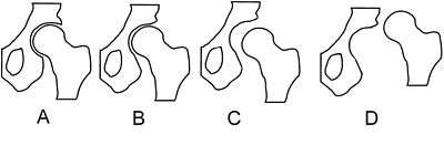
Crowe classification
In 1979 Dr. John F. Crowe[39] et al. proposed a classification to define the degree of malformation and dislocation. Grouped from least severe Crowe I dysplasia to most severe Crowe IV.[40] This classification is very useful for studying treatment results.
Rather than using the Wiberg angle because it makes it difficult to quantify the degree of dislocation they used 3 key elements to determine the degree of subluxation: A reference line at the lower rim of the "teardrop", junction between the femoral head and neck of the respective joint and the height of the pelvis (vertical measurement). They studied anteroposterior pelvic x-rays and drew horizontal lines through the lower rim of a feature called "teardrop". The distance between this line and the middle lines of the junction between femur head and neck gave them a measure of the degree of femur head subluxation. They further established that a "normal" diameter of the femur head measures 20% of the height of the pelvis. If the middle line of the neck-head junction was more than 10% of the pelvis height above the reference line they considered the joint to be more than 50% dislocated.
The following types resulted:
| Class | Description | Dislocation |
| Crowe I | Femur and acetabulum show minimal abnormal development. | Less than 50% dislocation |
| Crowe II | The acetabulum shows abnormal development. | 50% to 75% dislocation |
| Crowe III | The acetabula is developed without a roof. A false acetabulum develops opposite the dislocated femur head position. The joint is fully dislocated. | 75% to 100% dislocation |
| Crowe IV | The acetabulum is insufficiently developed. Since the femur is positioned high up on the pelvis this class is also known as "high hip dislocation". | 100% dislocation |
Treatment
Hip dysplasia presents a nearly perfect equilibrium between the arthritis, movement/mobility problems and pain associated with the developmental malformation, and the arthritis, movement/mobility problems and pain that are, as often as not in moderate to severe cases, inflicted by the treatment itself.
However, given the very real possibility of a limp, constant and/or debilitating pain, complicated treatment and impaired mobility later in life, careful developmental monitoring is indicated and early intervention is often the result. The worst possible consequence of non treatment is developing early arthritis, sometimes even during teenage years. All treatment aims to delay the onset of arthritis, but no treatment is fully successful in avoiding it; and, all available treatments bear the risk of inflicting equivalent damage. Most unfortunately, studies have as yet been unable to find a method of predicting outcomes in either the surgical/orthopedic treatment of the condition in infants and young children, or the surgical treatment of these early treatments' negative outcomes later in life (such as arthritis, avascular necrosis, trochanteric bursitis, and bone spurs of up to 2 cm just medial of the gluteus maximus insertion point on the greater trochanter due to excessive friction).
Harnesses, casts, and traction
Early hip dysplasia can often be treated using a Pavlik harness[41] (see photograph) or the Frejka pillow/splint[42] in the first year of life with usually normal results. Complications can occur when using the Pavlik Harness. Cases of Femoral Nerve Palsy[43] and Avascular Necrosis of the femoral head have been reported with the use of the Pavlik harness,[44] but whether these cases were due to improper application of the device or a complication encountered in the course of the disorder remains unresolved. Complications arise mainly because the sheet of the iliopsoas muscle pushes circumflex artery against the neck of the femur and decreases blood flow to the femoral head, so the Frejka pillow is not indicated in all the forms of the developmental dysplasia of the hip.
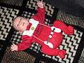 baby wearing a Bock harness
 Diagram of Pavlik harness
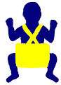 Diagram of Frejka pillow
 Traction
Other devices employed include the spica cast,[45] particularly following surgical closed reduction, open reduction, or osteotomy in babies and young children. Traction is sometimes used in the weeks leading up to a surgery to help stretch ligaments in the hip joint, although its use is controversial and varies amongst physicians.[46]
Surgery
In older children the adductor and iliopsoas muscles may have to be treated surgically because they adapt to the dislocated joint position (contracture). Braces and splints are often used following either of these methods to continue treatment. Although some children "outgrow" untreated mild hip dysplasia[37] and some forms of untreated dysplasia cause little or no impairment of quality of life, studies have as yet been unable to find a method of predicting outcomes. On the other hand, it has often been documented that starting treatment late leads to complications and ends in poor results.
Hip replacement and osteotomy
Hip dysplasia is often cited as causing osteoarthritis of the hip at a comparatively young age. Dislocated load bearing surfaces lead to increased and unusual wear. Although there are studies that contradict these findings. (see [47][48]) Peri-acetabular osteotomy (PAO) surgery can be used to realign the hip joint in some adolescents and adults. Subsequent treatment with total hip arthroplasty (hip replacement) is complicated by a need for revision surgery (replacing the artificial joint) owing to skeletal changes as the body matures, loosening/wear or bone resorption. Hip resurfacing is another option for correcting hip dysplasia in adults. It is a type of hip replacement that preserves more bone, and may work for younger hip dysplasia patients.[49]
Osteotomies are either used in conjunction with arthroplasty or by themselves to correct misalignment.
Epidemiology
Determining the incidence can be difficult.[50][51] In addition there is a wide margin in diagnostic results. A German study comparing two methods resulted in twice the usual rate for one method.
An instability rate of 1:60 has been described, though this rate drops to 1:240 at one week.[52] The condition is eight times more frequent in females than in males.[7]
History
The Frejka Pillow Splint was named after Dr. Bedrich Frejka (1890-1972), a Czech Orthopedic Surgeon. The Pavlik Harness was named after Dr. Arnold Pavlik (1902-1962), also a Czech Orthopedic Surgeon.
Society and culture
In the television program "ER," Kerry Weaver uses a crutch owing to congenital hip dysplasia. In season 12, she undergoes a hip replacement to cure her dysplasia when her previously untreated joint worsens.[53]
Other animals
In dogs, hip dysplasia is an abnormal formation of the hip socket that, in its more severe form, can eventually cause crippling lameness and painful arthritis of the joints. It is a genetic (polygenic) trait that is affected by environmental factors. It can be found in many animals, but is most commonly associated with dogs, and is common in many dog breeds, particularly the larger breeds.
Hip dysplasia is one of the most studied veterinary conditions in dogs, and the most common single cause of arthritis of the hips.[54]
Research
One avenue of research is using stem cells. They are applied in grafting (bone grafting) or by seeding porous arthroplasty prosthesis with autologous fibroblasts or chondrocyte progenitor cells to assist in firmly anchoring the artificial material in the bone bed.
References
- ↑ "Your Orthopaedic Connection: Developmental Dysplasia of the Hip".
- ↑ "Definition: congenital dysplasia of the hip from Online Medical Dictionary".
- ↑ Jacobsen S, Rømer L, Søballe K (2006). "The other hip in unilateral hip dysplasia". Clin. Orthop. Relat. Res. 446: 239–46. doi:10.1097/01.blo.0000201151.91206.50. PMID 16721954.
- ↑ Wiberg G. Studies of acetabular and congenital subluxation of the hip joint with special reference to complication of osteoarthritis Acta Chir Scand 1939, 83(Suppl. 58)
- ↑ Cilliers HJ, Beighton P (1990). "Beukes familial hip dysplasia: an autosomal dominant entity". Am. J. Med. Genet. 36 (4): 386–90. doi:10.1002/ajmg.1320360403. PMID 2389793.
- ↑ Lynn T Staheli, Fundamentals of Pediatric Orthopedics, p 13
- 1 2 Clinical Practice Guideline: Early Detection of Developmental Dysplasia of the Hip - Committee on Quality Improvement, Subcommittee on Developmental Dysplasia of the Hip 105 (4): 896 - AAP Policy
- ↑ Forst J, Forst C, Forst R, Heller KD (1997). "Pathogenetic relevance of the pregnancy hormone relaxin to inborn hip instability". Arch Orthop Trauma Surg. 116 (4): 209–12. doi:10.1007/BF00393711. PMID 9128773.
- ↑ CHSC01
- ↑ The occurrence of hip joint dislocation in early Lappic populations of Norway, Per Holck, Anthropological Department, Anatomical Institute, Box 1105 Blindern, N-0317 Oslo 3, Norway
- ↑ Forsdahl A (2000). "[A physician from Finnmark who pointed out the significance of heredity in congenital hip dysplasia]". Tidsskr. Nor. Laegeforen. (in Norwegian). 120 (22): 2672–3. PMID 11077514.
- ↑ Mabuchi A, Nakamura S, Takatori Y, Ikegawa S (2006). "Familial osteoarthritis of the hip joint associated with acetabular dysplasia maps to chromosome 13q". Am. J. Hum. Genet. 79 (1): 163–8. doi:10.1086/505088. PMC 1474113
 . PMID 16773577.
 - ↑ AJHG - Autosomal Dominant (Beukes) Premature Degenerative Osteoarthropathy of the Hip Joint Maps to an 11-cM Region on Chromosome 4q35
- ↑ Mahan ST, Kasser JR (2008). "Does swaddling influence developmental dysplasia of the hip?". Pediatrics. 121 (1): 177–8. doi:10.1542/peds.2007-1618. PMID 18166571.
- ↑ "Baby Carriers, Seats, & Other Equipment - International Hip Dysplasia Institute".
- ↑ "Hip-Healthy Swaddling - International Hip Dysplasia Institute".
- ↑ "Causes of Developmental Dysplasia of the Hip - International Hip Dysplasia Institute".
- ↑ "Developmental Dislocation of the Hip - Wheeless' Textbook of Orthopaedics".
- ↑ "HipDysplasia - Newborn Nursery at LPCH - Stanford University School of Medicine".
- ↑ Kamath S, Bramley D (2005). "Is 'clicky hip' a risk factor in developmental dysplasia of the hip?". Scott Med J. 50 (2): 56–8. PMID 15977515.
- ↑ "Newborn Screening and Prevention - International Hip Dysplasia Institute".
- ↑ "Hip Clicks and Hip Dysplasia - International Hip Dysplasia Institute".
- ↑ "Physical Examination of Infants - International Hip Dysplasia Institute".
- ↑ "Ultrasound Detection of DDH - International Hip Dysplasia Institute".
- ↑ "X-Ray Screening for Developmental Dysplasia of the Hip - International Hip Dysplasia Institute".
- ↑ "Shenton's line (www.whonamedit.com)".
- ↑ "Radiographic features: DDH". Weeless' Textbook of Orthopaedics.
- ↑ "Asymmetry Symptoms of DDH".
- ↑ "Physician Newborn Screening and Prevention - International Hip Dysplasia Institute".
- ↑ Harris Hip Score - Orthopaedic Scores
- ↑ Wylde V, Learmonth ID, Cavendish VJ (2005). "The Oxford hip score: the patient's perspective". Health Qual Life Outcomes. 3: 66. doi:10.1186/1477-7525-3-66. PMC 1283979. PMID 16259627.
- ↑ Aguilar CM, Neumayr LD, Eggleston BE, et al. (2005). "Clinical evaluation of avascular necrosis in patients with sickle cell disease: Children's Hospital Oakland Hip Evaluation Scale--a modification of the Harris Hip Score". Arch Phys Med Rehabil. 86 (7): 1369–75. doi:10.1016/j.apmr.2005.01.008. PMID 16003666.
- ↑ Betsy Miller, The Parents' Guide to Hip Dysplasia, p 19.
- ↑ Sutherland, Denise; West, Sophie (2011). A Guide for Adults with Hip Dysplasia. pp. 7, 21–23. ISBN 978-0-9872152-0-8.
- ↑ "Developmental Dysplasia of the Hip - October 15, 2006 -- American Family Physician".
- ↑ Dr. Rose's Peripheral Brain-DEVELOPMENTAL DYSPLASIA OF THE HIP
- 1 2 Screening for Developmental Dysplasia of the Hip - July 1999 - American Academy of Family Physicians
- ↑ "eMedicine - Developmental Dysplasia of the Hip : Article by James McCarthy, MD, FAAOS".
- ↑ http://www.onsmd.com/Jon-F-Crowe-MD
- ↑ Crowe JF, Mani VJ, Ranawat CS (1979). "Total hip replacement in congenital dislocation and dysplasia of the hip". J Bone Joint Surg Am. 61 (1): 15–23. PMID 365863.
- ↑ "Pavlik Harness".
- ↑ Czubak J, Piontek T, Niciejewski K, Magnowski P, Majek M, Płończak M (2004). "Retrospective analysis of the non-surgical treatment of developmental dysplasia of the hip using Pavlik harness and Fredjka pillow: comparison of both methods". Ortop Traumatol Rehabil. 6 (1): 9–13. PMID 17676003.
- ↑ "Femoral Nerve Palsy".
- ↑ Nakamura J, Kamegaya M, Saisu T, Someya M, Koizumi W, Moriya H (2007). "Treatment for developmental dysplasia of the hip using the Pavlik harness: long-term results". J Bone Joint Surg Br. 89 (2): 230–5. doi:10.1302/0301-620X.89B2.18057. PMID 17322441.
- ↑ "Hip Spica Cast for Developmental Dysplasia of the Hip".
- ↑ "Traction with DDH Treatment".
- ↑ Lau EM, Lin F, Lam D, Silman A, Croft P (1995). "Hip osteoarthritis and dysplasia in Chinese men". Ann. Rheum. Dis. 54 (12): 965–9. doi:10.1136/ard.54.12.965. PMC 1010061. PMID 8546528.
- ↑ Lievense AM, Bierma-Zeinstra SM, Verhagen AP, Verhaar JA, Koes BW (2004). "Influence of hip dysplasia on the development of osteoarthritis of the hip". Ann. Rheum. Dis. 63 (6): 621–6. doi:10.1136/ard.2003.009860. PMC 1755018. PMID 15140766.
- ↑ Sutherland, Denise; West, Sophie (2011). A Guide for Adults with Hip Dysplasia. pp. 56–59. ISBN 978-0-9872152-0-8.
- ↑ Bialik V, Bialik GM, Blazer S, Sujov P, Wiener F, Berant M (1999). "Developmental dysplasia of the hip: a new approach to incidence". Pediatrics. 103 (1): 93–9. doi:10.1542/peds.103.1.93. PMID 9917445.
- ↑ Kokavec M, Bialik V (2007). "Developmental dysplasia of the hip. Prevention and real incidence". Bratisl Lek Listy. 108 (6): 251–4. PMID 17972535.
- ↑ "UNSW Embryology- Musculoskeletal System - Abnormalities".
- ↑ "ER - Out on a Limb (2006)". Retrieved 31 August 2012.
- ↑ Workingdogs.com. "Canine hip dysplasia". Workingdogs.com. Retrieved 2013-08-18.
External links
- International Hip Dysplasia Institute (IHDI)
- Online orthopedic textbook
- Children's Hospital Central California - Developmental Dysplasia of the Hip (DDH)
- United Kingdom Support group for DDH, Clubfoot and other lower limb disorders
- United Kingdom Support Links for Hip Dysplasia