Dental composite
Dental composite resins are types of synthetic resins which are used in dentistry as restorative material or adhesives. Synthetic resins evolved as restorative materials since they were insoluble, aesthetic, insensitive to dehydration, easy to manipulate and reasonably inexpensive. Composite resins are most commonly composed of Bis-GMA and other dimethacrylate monomers (TEGMA, UDMA, HDDMA), a filler material such as silica and in most current applications, a photoinitiator. Dimethylglyoxime is also commonly added to achieve certain physical properties such as flow ability. Further tailoring of physical properties is achieved by formulating unique concentrations of each constituent.
Many studies have compared the longevity of composite restorations to the longevity of silver-mercury amalgam restorations. Depending on the skill of the dentist, patient characteristics and the type and location of damage, composite restorations can have similar longevity to amalgam restorations. (See Longevity and clinical performance.) In comparison to amalgam, the aesthetics of composite restorations are far superior.
History of use
Initially, composite restorations in dentistry were very prone to leakage and breakage due to weak compressive strength. In the 1990s and 2000s, composites were greatly improved and have a compression strength sufficient for use in posterior teeth.
Method and clinical application
Today's composite resins have low polymerization shrinkage and low coefficients of thermal shrinkage, which allows them to be placed in bulk while maintaining good adaptation to cavity walls. The placement of composite requires meticulous attention to procedure or it may fail prematurely. The tooth must be kept perfectly dry during placement or the resin will likely fail to adhere to the tooth. Composites are placed while still in a soft, dough-like state, but when exposed to light of a certain blue wavelength (typically 470 nm[1]), they polymerize and harden into the solid filling (for more information, see Light activated resin). It is challenging to harden all of the composite, since the light often does not penetrate more than 2–3 mm into the composite. If too thick an amount of composite is placed in the tooth, the composite will remain partially soft, and this soft unpolymerized composite could ultimately lead to leaching of free monomers with potential toxicity and/or leakage of the bonded joint leading to recurring dental pathology. The dentist should place composite in a deep filling in numerous increments, curing each 2–3 mm section fully before adding the next. In addition, the clinician must be careful to adjust the bite of the composite filling, which can be tricky to do. If the filling is too high, even by a subtle amount, that could lead to chewing sensitivity on the tooth. A properly placed composite is comfortable, aesthetically pleasing, strong and durable, and could last 10 years or more. (By most North American insurance companies 2 years minimum)
The most desirable finish surface for a composite resin can be provided by aluminum oxide disks. Classically, Class III composite preparations were required to have retention points placed entirely in dentin. A syringe was used for placing composite resin because the possibility of trapping air in a restoration was minimized. Modern techniques vary, but conventional wisdom states that because there have been great increases in bonding strength due to the use of dentin primers in the late 1990s, physical retention is not needed except for the most extreme of cases. Primers allow the dentin's collagen fibers to be "sandwiched" into the resin, resulting in a superior physical and chemical bond of the filling to the tooth. Indeed, composite usage was highly controversial in the dental field until primer technology was standardized in the mid to late 1990s. The enamel margin of a composite resin preparation should be beveled in order to improve aesthetics and expose the ends of the enamel rods for acid attack. The correct technique of enamel etching prior to placement of a composite resin restoration includes etching with 30%-50% phosphoric acid and rinsing thoroughly with water and drying with air only. In preparing a cavity for restoration with composite resin combined with an acid etch technique, all enamel cavosurface angles should be obtuse angles. Contraindications for composite include varnish and zinc oxide-eugenol. Composite resins for Class II restorations were not indicated because of excessive occlusal wear in the 1980s and early 1990s. Modern bonding techniques and the increasing unpopularity of amalgam filling material have made composites more attractive for Class II restorations. Opinions vary, but composite is regarded as having adequate longevity and wear characteristics to be used for permanent Class II restorations. Whether composite materials last as long or has the leakage and sensitivity properties when compared to Class II amalgam restorations was described as a matter of debate in 2008.[2]
Composition

As with other composite materials, a dental composite typically consists of a resin-based oligomer matrix, such as a bisphenol A-glycidyl methacrylate (BISGMA), urethane dimethacrylate (UDMA) or [(semi-crystalline polyceram)] (PEX), and an inorganic filler such as silicon dioxide (silica). Compositions vary widely, with proprietary mixes of resins forming the matrix, as well as engineered filler glasses and glass ceramics. The filler gives the composite wear resistance and translucency. A coupling agent such as silane is used to enhance the bond between these two components. An initiator package (such as: camphorquinone (CQ), phenylpropanedione (PPD) or lucirin (TPO)) begins the polymerization reaction of the resins when external energy (light/heat, etc.) is applied. A catalyst package can control its speed.
Advantages
Advantages of composites:
- Aesthetics: The main advantage of a direct dental composite over traditional materials such as amalgam is improved aesthetics. Composites can be in a wide range of tooth colors allowing near invisible restoration of teeth. Composite fillings can be closely matched to the color of existing teeth.
- Bonding to tooth structure: Composite fillings micro-mechanically bond to tooth structure. This strengthens the tooth's structure and restores its original physical integrity. The discovery of acid etching (producing enamel irregularities ranging from 5-30 micrometers in depth) of teeth to allow a micro-mechanical bond to the tooth allows good adhesion of the restoration to the tooth. Very high bond strengths to tooth structure, both enamel and dentin, can be achieved with the current generation of dentin bonding agents.
- Tooth-sparing preparation: The fact that composite fillings are glued (bonded) to the tooth means that unlike amalgam fillings, there is no need for the dentist to create retentive features destroying healthy tooth. Unlike amalgam, which just fills a hole and relies on the geometry of the hole to retain the filling, composite materials are bonded to the tooth. In order to achieve the necessary geometry to retain an amalgam filling, the dentist may need to drill out a significant amount of healthy tooth material. In the case of a composite restoration, the geometry of the hole (or "box") is less important because a composite filling bonds to the tooth. Therefore less healthy tooth needs to be removed for a composite restoration.
- Less-costly and more conservative alternative to dental crowns: In some situations, a composite restoration may be offered as a less-expensive (though possibly less durable) alternative to a dental crown, which can be a very expensive treatment. Installation of a dental crown usually requires removal of significant healthy tooth material so the crown can fit over or into the natural tooth. Composite restoration conserves more of the natural tooth.
- Alternative to tooth removal: Because a composite restoration bonds to the tooth and can restore the original physical integrity of a damaged or decayed tooth, in some cases composite restoration can preserve a tooth that might not be salvageable with amalgam restoration. For example, depending on the location and extent of decay, it might not be possible to create a void (a "box") of the geometry necessary to retain an amalgam filling.
- Versatility: Composite fillings can be used to repair chipped, broken or worn teeth[3] which would not be repairable using amalgam fillings.
- Repairability: In many cases of minor damage to a composite filling, the damage can be easily repaired by adding additional composite. An amalgam filling might require complete replacement.
- Reduced quantity of mercury released to the environment: Composites avoid mercury environmental contamination associated with dentistry. When amalgam fillings are drilled for height adjustment, repair or replacement, some mercury-containing amalgam is inevitably washed down drains. (See Dental amalgam controversy - Environmental impact) When amalgam fillings are prepared by dentists, improperly disposed excess material may enter landfills or be incinerated. Cremation of bodies containing amalgam fillings releases mercury into the environment. (See Dental amalgam controversy - Cremation)
- Reduced mercury exposure for dentists: Preparing new amalgam fillings and drilling into existing amalgam fillings exposes dentists to mercury vapor. Use of composite fillings avoids this risk, unless the procedure also involves removing an existing amalgam filling. A review article found studies indicating that dental work involving mercury may be an occupational hazard with respect to reproductive processes, glioblastoma (brain cancer), renal function changes, allergies and immunotoxicological effects.[4] (See Dental amalgam controversy - Health effects for dentists)
- Lack of corrosion: Although corrosion is no longer a major problem with amalgam fillings, resin composites do not corrode at all. (Low-copper amalgams, prevalent before 1963, were more subject to corrosion than modern high-copper amalgams.[5] )
Disadvantages
- Composite shrinkage and secondary caries: In the past, composite resins suffered significant shrinkage during curing, which led to inferior bonding interface.[6] Shrinkage permits microleakage, which, if not caught early, can cause secondary caries (subsequent decay), the most significant dental disadvantage of composite restoration. In a study of 1,748 restorations, risk of secondary caries in the composite group was 3.5 times risk of secondary caries in the amalgam group.[7] Good dental hygiene and regular checkups can mitigate this disadvantage. Most current microhybrid and nanohybrid composites have a polymerization shrinkage that ranges from 2% to 3.5%. Composite shrinkage can be reduced by altering the molecular and bulk composition of the resin.[8] In the field of dental restorative materials, reduction of composite shrinkage has been achieved with some success.[2] Among the newest materials, silorane resin exhibits lower polymerization shrinkage, compared to the dimethacrylates.[2]
- Durability: In some situations, composite fillings may not last as long as amalgam fillings under the pressure of chewing, particularly if used for large cavities. (See Longevity and clinical performance, below.)
- Chipping: Composite materials can chip off the tooth.
- Skill and training required: Successful outcomes in direct composite fillings is related to the skills of the practitioner and technique of placement,.[2] For example, a rubber dam is rated to be important to achieve low fracture rates and a longevity similar to amalgam in the more demanding proximal Class II cavities.[9]
- Need to keep working area in mouth completely dry: The prepared tooth must be completely dry (free of saliva and blood) when the resin material is being applied and cured. Posterior teeth (molars) are difficult to keep dry. Keeping the prepared tooth completely dry can also be difficult for any work involving treatment of cavities below the gumline.[10]
- Time and expense: Due to the sometimes complicated application procedures and the need to keep the prepared tooth absolutely dry, composite restorations may take up to 20 minutes longer than equivalent amalgam restorations.[10] Longer time in the dental chair may test the patience of children, making the procedure more difficult for the dentist. Due to the longer time involved, the fee charged by a dentist for a composite restoration may be higher than for an amalgam restoration.[3]
- Limited insurance coverage: Some dental insurance plans may provide reimbursement for composite restoration only on front teeth where amalgam restorations would be particularly objectionable on cosmetic grounds. Thus, patients may be required to pay the entire charge for composite restorations on posterior teeth. For example one dental insurer states that most of their plans will pay for resin (i.e. composite) fillings only "on the teeth where their cosmetic benefit is critical: the six front teeth (incisors and cuspids) and on the facial (cheek side) surfaces of the next two teeth (bicuspids)."[10] Even if charges are paid by private insurance or government programs, the higher cost is incorporated in dental insurance premiums or tax rates.
Direct dental composites
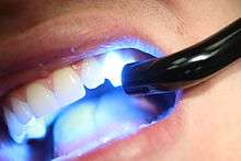
Direct dental composites are placed by the dentist in a clinical setting. Polymerization is accomplished typically with a hand held curing light that emits specific wavelengths keyed to the initiator and catalyst packages involved. When using a curing light, the light should be held as close to the resin surface as possible, a shield should be placed between the light tip and the operator's eyes. Curing time should be increased for darker resin shades. Light cured resins provide denser restoration than self-cured resins because no mixing is required that might introduce air bubble porosity.
Direct dental composites can be used for:
- Filling cavity preparations
- Filling gaps (diastemas) between teeth using a shell-like veneer or
- Minor reshaping of teeth
- Partial crowns on single teeth
Indirect dental composites
Indirect composite is cured outside the mouth, in a processing unit that is capable of delivering higher intensities and levels of energy than handheld lights can. Indirect composites can have higher filler levels, are cured for longer times and curing shrinkage can be handled in a better way. As a result, they are less prone to shrinkage stress and marginal gaps[11] and have higher levels and depths of cure than direct composites. For example, an entire crown can be cured in a single process cycle in an extra-oral curing unit, compared to a millimeter layer of a filling.
As a result, full crowns and even bridges (replacing multiple teeth) can be fabricated with these systems.
Indirect dental composites can be used for:
- Filling cavities in teeth, as fillings, inlays and/or onlays
- Filling gaps (diastemas) between teeth using a shell-like veneer or
- Reshaping of teeth
- Full or partial crowns on single teeth
- Bridges spanning 2-3 teeth
A stronger, tougher and more durable product is expected in principle. But in the case of inlays, not all clinical long-term-studies detect this advantage in clinical practice (see below).
Longevity and clinical performance
Direct composite vs amalgam
Clinical survival of composite restorations placed in posterior teeth are in the range of amalgam restorations, with some studies seeing a slightly lower[12] or slightly higher[13] survival time compared to amalgam restorations. Improvements in composite technology and application technique make composites a very good alternative to amalgam, while use in large restorations and in cusp capping situations is still debated.[2]
According to a 2012 review article by Demarco et al. covering 34 relevant clinical studies, "90% of the studies indicated that annual failure rates between 1% and 3% can be achieved with Class I and II posterior [rear tooth] composite restorations depending on the definition of failure, and on several factors such as tooth type and location, operator [dentist], and socioeconomic, demographic, and behavioral elements." [14] This compares to a 3% mean annual failure rate reported in a 2004 review article by Manhart et al. for amalgam restorations in posterior stress-bearing cavities.[15][16]
The Demarco review found that the main reasons cited for failure of posterior composite restorations are secondary caries (i.e. cavities which develop subsequent to the restoration), fracture, and patient behavior, notably bruxism (grinding/clenching.) Causes of failure for amalgam restorations reported in the Manhart et al.review also include secondary caries, fracture (of the amalgam and/or the tooth), as well as cervical overhang and marginal ditching.[16] The Demarco et al. review of composite restoration studies noted that patient factors affect longevity of restorations: Compared to patients with generally good dental health, patients with poorer dental health (possibly due to poor dental hygiene, diet, genetics, frequency of dental checkups, etc.) experience higher rates of failure of composite restorations due to subsequent decay.[17] Socioeconomic factors also play a role: "People who had always lived in the poorest stratus [sic][stratum?] of the population had more restoration failures than those who lived in the richest layer."[14]
The definition of failure applied in clinical studies may affect the reported statistics. Demarco et al note: "Failed restorations or restorations presenting small defects are routinely treated by replacement by most clinicians. Because of this, for many years, the replacement of defective restorations has been reported as the most common treatment in general dental practice..."[14] Demarco et al observe that when both repaired and replaced restorations were classified as failures in one study, the Annual Failure Rate was 1.9%. However, when repaired restorations were reclassified as successes instead of failures, the AFR decreased to 0.7%. Reclassifying repairable minor defects as successes rather than failures is justifiable: "When a restoration is replaced, a significant amount of sound tooth structure is removed and the preparation [i.e. hole] is enlarged".[18][19] Applying the narrower definition of failure would improve the reported longevity of composite restorations: Composite restorations can often be easily repaired or extended without drilling out and replacing the entire filling. Resin composites will adhere to the tooth and to undamaged prior composite material. In contrast, amalgam fillings are held in place by the shape of the void being filled rather than by adhesion. This means that it is often necessary to drill out and replace an entire amalgam restoration rather than add to the remaining amalgam.
Direct vs indirect composites
It might be expected that the costlier indirect technique leads to a higher clinical performance, however this is not seen in all studies. A study conducted over the course of 11 years reports similar failure rates of direct composite fillings and indirect composite inlays.[11] Another study concludes that although there is a lower failure rate of composite inlays it would be insignificant and anyway too small to justify the additional effort of the indirect technique.[20] Also in the case of ceramic inlays a significantly higher survival rate compared to composite direct fillings can not be detected.[21]
In general, a clear superiority of tooth colored inlays over composite direct fillings could not be established by current review literature (as of 2013).[22] [23] [24]
See also
References
- ↑ Rueggeberg, Frederick (2011). "State-of-the-art: Dental Photocuring - A review". Dental Materials. 27: 39–52. doi:10.1016/j.dental.2010.10.021.
- 1 2 3 4 5 Shenoy, A. (2008). "Is it the end of the road for dental amalgam? A critical review". Journal of Conservative Dentistry (
 ). 11 (3): 99–107. doi:10.4103/0972-0707.45247. PMC 2813106
 . PMID 20142895.
 - 1 2 "Dental Health and Tooth Fillings". WebMD. Retrieved 23 November 2013.
- ↑ Bjørklund G (1991). "Mercury in the dental office. Risk evaluation of the occupational environment in dental care (in Norwegian)". Tidsskr Nor Laegeforen. 111 (8): 948–951. PMID 2042211.
- ↑ Bharti, Ramesh. "Dental amalgam: An update". Retrieved 23 November 2013.
- ↑ Schneider LF, Cavalcante LM, Silikas N.; "Shrinkage Stresses Generated during Resin-Composite Applications: A Review" J Dent Biomech. 2010 doi:10.4061/2010/131630
- ↑ Bernardo, Mario. "Survival and reasons for failure of amalgam versus composite posterior restorations placed in a randomized clinical trial" (PDF). J Am Dent Assoc. p. 779. Retrieved 23 November 2013.
- ↑ For example, UltraSeal XT Plus uses Bis-GMA without dimethacrylate and was found to have a shrinkage of 5.63%, 30 minutes after curing. On the other hand, this same study found that Heliomolar, which uses Bis-GMA, UDMA and decandiol dimethacrylate, had a shrinkage of 2.00%, 30 minutes after curing.KLEVERLAAN, CJ; Feilzer, AJ (2005). "Polymerization shrinkage and contraction stress of dental resin composites". Dental Materials. 21 (12): 1150–7. doi:10.1016/j.dental.2005.02.004. PMID 16040118. Retrieved 2009-04-16.
- ↑ Heintze, S. D.; Rousson, V. (2012). "Clinical effectiveness of direct class II restorations - a meta-analysis". The journal of adhesive dentistry. 14 (5): 407–431. doi:10.3290/j.jad.a28390. PMID 23082310.
- 1 2 3 "Dental amalgam or resin composite fillings?". Delta Dental. Retrieved 23 November 2013.
- 1 2 Composite resin fillings and inlays. An 11-year evaluation.; U Pallesen, V Qvist; (2003) Clin Oral Invest 7:71–79 doi:10.1007/s00784-003-0201-z Conclusion:.." Considering the more invasive cavity preparation and the higher cost of restorations made by the inlay technique, this study indicates that resin fillings in most cases should be preferred over resin inlays."
- ↑ Bernardo, M.; Luis, H.; Martin, M. D.; Leroux, B. G.; Rue, T.; Leitão, J.; Derouen, T. A. (2007). "Survival and reasons for failure of amalgam versus composite posterior restorations placed in a randomized clinical trial". Journal of the American Dental Association (1939). 138 (6): 775–783. doi:10.14219/jada.archive.2007.0265. PMID 17545266.
- ↑ J. Manhart, H. Chen, G. Hamm, R. Hickel: Buonocore Memorial Lecture. Review of the clinical survival of direct and indirect restorations in posterior teeth of the permanent dentition. In: Operative dentistry. Band 29, Nummer 5, 2004 Sep-Oct, S. 481–508, ISSN 0361-7734. PMID 15470871. (Review).
- 1 2 3 Demarco FF1, Corrêa MB, Cenci MS, Moraes RR, Opdam NJ (2012). "Longevity of posterior composite restorations: not only a matter of materials". Dental Materials. 28 (1): 87–101. doi:10.1016/j.dental.2011.09.003. PMID 22192253.
- ↑ Manhart, J; Chen H; Hamm G; Hickel R (Sep–Oct 2004). "Buonocore Memorial Lecture. Review of the clinical survival of direct and indirect restorations in posterior teeth of the permanent dentition". Oper Dent. 29 (5): 481–508. PMID 15470871.
- 1 2 Shenoy, Arvind (Jul–Sep 2008). "Is it the end of the road for dental amalgam? A critical review". Journal of Conservative Dentistry. 11 (3): 99–107. doi:10.4103/0972-0707.45247. PMC 2813106. PMID 20142895.
- ↑ "Longevity of posterior composite restorations: Not only a matter of materials". p. 92.
- ↑ Moncada, G; Martin J; Fernandez E; Hempel MC; Mjor IA; Gordan VV (2009). "Sealing, refurbishment and repair of Class I and Class II defective restorations: a three-year clinical trial". J Am Dent Assoc. 140 (4): 425–32. doi:10.14219/jada.archive.2009.0191. PMID 19339531.
- ↑ Gordan, VV; Riley 3rd JL; Blaser PK; Mondragon E; Garvan CW; Mjor IA (2011). "Alternative treatments to replacement of defective amalgam restorations: results of a seven-year clinical study". J Am Dent Assoc. 142: 842–9. doi:10.14219/jada.archive.2011.0274.
- ↑ Direct resin composite inlays/onlays: an 11 year follow-up. JWV Van Dijken; (2000) J Dent 28:299–306; PMID 10785294
- ↑ Clinical evaluation of ceramic inlays compared to composite restorations.; (2009); RT Lange, P Pfeiffer; Oper Dent. May-Jun;34(3):263-72. doi:10.2341/08-95.
- ↑ Goldstein, G. R. (2010). "The Longevity of Direct and Indirect Posterior Restorations is Uncertain and may be Affected by a Number of Dentist-, Patient-, and Material-Related Factors". Journal of Evidence Based Dental Practice (Review Article). 10 (1): 30–31. doi:10.1016/j.jebdp.2009.11.015. PMID 20230962.
- ↑ Critchlow, S. (2012). "Ceramic materials have similar short term survival rates to other materials on posterior teeth". Evidence-Based Dentistry. 13 (2): 49. doi:10.1038/sj.ebd.6400860. PMID 22722415. Conclusions: "Ceramic materials perform as well as alternative restorative materials for use as inlay restorations. However, a lack of long-term data means that this conclusion can only be supported for periods up to one year for longevity.."
- ↑ Thordrup, M.; Isidor, F.; Hörsted-Bindslev, P. (2006). "A prospective clinical study of indirect and direct composite and ceramic inlays: Ten-year results". Quintessence international (Berlin, Germany : 1985). 37 (2): 139–144. PMID 16475376.
| Wikimedia Commons has media related to Composite Fillings. |
| Wikimedia Commons has media related to Polymerisation lamps. |