Lateralization of brain function
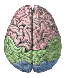
The lateralization of brain function refers to how some neural functions, or cognitive processes tend to be more dominant in one hemisphere than the other. The medial longitudinal fissure separates the human brain into two distinct cerebral hemispheres, connected by the corpus callosum. Although the macrostructure of the two hemispheres appears to be almost identical, different composition of neuronal networks allows for specialized function that is different in each hemisphere.[1]
Lateralization of brain structures is based on general trends expressed in healthy patients; however, there are numerous counterexamples to each generalization. Each human’s brain develops differently leading to unique lateralization in individuals. This is different from specialization as lateralization refers only to the function of one structure divided between two hemispheres. Specialization is much easier to observe as a trend since it has a stronger anthropological history.[2] The best example of an established lateralization is that of Broca's and Wernicke's Areas where both are often found exclusively on the left hemisphere. These areas frequently correspond to handedness however, meaning the localization of these areas is regularly found on the hemisphere corresponding to the dominant hand (anatomically on the opposite side). Function lateralization such as semantics, intonation, accentuation, prosody, etc. has since been called into question and largely been found to have a neuronal basis in both hemispheres.[3][4]
Interaction and Role
Theme
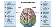
To get a basic understanding of this complex issue it is easiest to first consider the left (LHS) and right (RHS) hemispheres as distinct but interacting entities. These interactions come in the form of both excitatory and inhibitory signals crossing the corpus callosum and other hemispheric bridges.[5][6] As LHS and RHS each have unique interpretations of situations these signals allow for discussion and an ultimate decision to be made.[7] This interaction is called hemispheric rivalry.[8] This term is apt as both hemispheres are in conflict yet on the same team. In humans the reliance on both hemispheres is the basis of a number of functions including consciousness.[9]
The LHS can be simplified to better understand its role in this rivalry. The LHS is centered around action and is often the driving force behind risky behaviors. This hemisphere heavily relies upon emotional input leading it to make brash and uncalculated decisions. These decisions should not be thought of as ill-conceived, rather illogical and raw.[10]
Similarly the RHS can be brought into understanding by discussing a simple model of its role in the hemispheric interactions. The RHS can be thought of as the opposite of LHS as it relies primarily on critical thinking and calculations to reach its decisions.[11] As such the conclusions reached by the RHS often result in avoidance of risk taking behaviors and overall inaction.[12]
When viewed as action (LHS) and inaction (RHS) the anthropological development of these structures becomes elementary and logical. In environments of scarcity, like those faced by non-human animals, taking risks is the foundational approach to survival. In scarcity it is far more likely to die of starvation than to damaging stimuli from hostile animals or situations. However, in environments of abundance, as humans have observed, it is far more likely to die to damaging stimuli than of starvation. As such the anthropological development of first LHS then much later RHS is understandable.[13]
This also brings clarity to differences observed in modern human brains between environments. In areas of prosperity, where warmth, food, and basic needs for survival are abundant RHS domination is prevalent. Unsurprisingly, in areas of scarcity where cold and limited food are concerns LHS domination is prevalent. This phenomenon has been recorded numerous times when examining LHS dominant cultures, such as those of the Arctic, to RHS dominant cultures, like Africa.[14] Similarly, studies of animals such as the Chickadee have shown similar results. Yukon born chickadees have LHS dominance as compared to Texas born chickadees with RHS dominance. In exchanging Yukon borns for Texas borns it was shown that the action reliant Yukon birds consistently became the alpha of their new environment whereas inaction reliant Texan birds died shortly after their arrival in the Yukon.
When speaking of dominance it is important to recognize that each hemisphere continues to function semi-independently but their interactions become dominated by one side. That is, each hemisphere always provides its input to the decision making process but one is drowned out by the other. This occurs as individual decisions are made that biologically alter the state of the brain, changing the weight each hemisphere carries in their rivalry. As a choice of activity or inactivity is made it influences how effectively one hemisphere can inhibit the other and simultaneously teaches the now less effective inhibiting hemisphere to provide more excitatory signals with more frequency. For example: if a child eats a cookie their LHS has successfully inhibited their RHS changing the power dynamic as to make LHS ever so slightly more dominant and RHS more submissive in their rivalry. The reverse is also true should the child not eat the cookie.[15][16]
A lifetime of these anatomically changing decisions, paired with environmental circumstances as discussed above, dictate the structure and plasticity of the human brain. In cases such as common suburban living the LHS has less distinct neural networks and appears significantly blander than the RHS. The opposite asymmetry is observed in individuals such as violent offenders whose LHS is more distinct and pronounced than the RHS. The highest degree of symmetry between the hemispheres has been studied in veteran gang members. These individuals display an amazing propensity to act in extremely risky ways, yet inhibit themselves in the face of provocation to survive.[17][18][19]
Specifics
In gaining a fuller understanding of the interaction and function of cerebral hemispheres one must trace neural networks. The basis of the correlation of LHS with action and emotion is its connectivity with specialized parts throughout both hemispheres that play a role in those behaviors. Most notably LHS has been shown to have integral connections to insula and amygdala. Similarly, the association of RHS with inaction and calculation is tied to its extensive networks connecting to the anterior cingulate, orbitofrontal and prefrontal cortices.[20] These specialized areas are implicated in the calculative and inhibitory processes of inaction and those related to delayed gratification.[21][22] In studies looking at individuals with damaged LHS invariably the temporal lobe, insula and amygdala also sustained connectivity damage.[23] Limiting the connectivity of such areas seriously limits the effectiveness by which they operate. This means that although logistically the LHS is not the ‘emotional’ hemisphere its connectivity with emotion related areas is crucial to their functioning.[24] This same process implicates the RHS as the ‘calculative’ hemisphere, though logistically it is not.[25][26]
The role of hemispheric rivalry has become a major talking point for those studying the generation of consciousness within the brain. Some believe the brain’s state of conflict is integrally linked to intelligence and genuine free will.[27]
Failures of lateralization
Lateralization as a concept fails because the brain consistently updates, consolidates, shifts information between the hemispheres.[28] In patients with damage to the LHS, functions traditionally associated with LHS were found predominantly in the RHS, most notably semantic choice.[29] These results hold for those with RHS damage who show a similar amount of functionality in specific motor skills via new neural connections developed in the LHS after RHS damage.[30] Amazingly these shifts continue to maximize brain potential even after extensive damage.[31] In studies where patients incur LHS damage and functionality is shifted to RHS, damage of the same magnitude to the RHS causes RHS functionality to become split once more. This effect has been shown on a neuronal level as the stimulation of specific neurons in the RHS causes similar responses in both hemispheres as neuron clusters on each side enlarge to compensate for the initial stimulation.[32] This points to a sort of equilibrium observed by the hemispheres.[33]
The shift of info and neuron functionality between hemispheres should not be surprising, however, as it has been observed in individuals who have lost a sense. In these individuals, neurons, even those that are specialized, are semi-repurposed to compensate for the loss. For example: those who become blind after years of vision are able to repurpose specialized sections of their brain to have increased visualization and nonspecialization to aid in the efficiency of their other senses.
History of research on lateralization
Broca
One of the first indications of brain function lateralization resulted from the research of French physician Pierre Paul Broca, in 1861. His research involved the male patient nicknamed "Tan", who suffered a speech deficit (aphasia); "tan" was one of the few words he could articulate, hence his nickname. In Tan's autopsy, Broca determined he had a syphilitic lesion in the left cerebral hemisphere. This left frontal lobe brain area (Broca's area) is an important speech production region. The motor aspects of speech production deficits caused by damage to Broca’s area are known as expressive aphasia. In clinical assessment of this aphasia, it is noted that the patient cannot clearly articulate the language being employed.
Wernicke
German physician Karl Wernicke continued in the vein of Broca's research by studying language deficits unlike expressive aphasia. Wernicke noted that not every deficit was in speech production; some were linguistic. He found that damage to the left posterior, superior temporal gyrus (Wernicke's area) caused language comprehension deficits rather than speech production deficits, a syndrome known as receptive aphasia.
Advance in imaging technique
These seminal works on hemispheric specialization were done on patients or postmortem brains, raising questions about the potential impact of pathology on the research findings. New methods permit the in vivo comparison of the hemispheres in healthy subjects. Particularly, magnetic resonance imaging (MRI) and positron emission tomography (PET) are important because of their high spatial resolution and ability to image subcortical brain structures.
Movement and sensation
In the 1940s, neurosurgeon Wilder Penfield and his neurologist colleague Herbert Jasper developed a technique of brain mapping to help reduce side effects caused by surgery to treat epilepsy. They stimulated motor and somatosensory cortices of the brain with small electrical currents to activate discrete brain regions. They found that stimulation of one hemisphere's motor cortex produces muscle contraction on the opposite side of the body. Furthermore, the functional map of the motor and sensory cortices is fairly consistent from person to person; Penfield and Jasper's famous pictures of the motor and sensory homunculi were the result.
Split-brain patients
Research by Michael Gazzaniga and Roger Wolcott Sperry in the 1960s on split-brain patients led to an even greater understanding of functional laterality. Split-brain patients are patients who have undergone corpus callosotomy (usually as a treatment for severe epilepsy), a severing of a large part of the corpus callosum. The corpus callosum connects the two hemispheres of the brain and allows them to communicate. When these connections are cut, the two halves of the brain have a reduced capacity to communicate with each other. This led to many interesting behavioral phenomena that allowed Gazzaniga and Sperry to study the contributions of each hemisphere to various cognitive and perceptual processes. One of their main findings was that the right hemisphere was capable of rudimentary language processing, but often has no lexical or grammatical abilities.[34] Eran Zaidel also studied such patients and found some evidence for the right hemisphere having at least some syntactic ability.
Language is primarily localized in the left hemisphere. One of the experiments carried out by Gazzaniga involved a split-brain patient sitting in front of a computer screen while having words and images presented on either side of the screen and the visual stimuli would go to either the right or left visual field, and thus the left or right brain, respectively. It was observed that if a patient was presented with an image to his left visual field (right brain), he would report not seeing anything. If he was able to feel around for certain objects, he could accurately pick out the correct object, despite not having the ability to verbalize what he saw. This led to confirmation that the left brain is localized for language while the right brain does not have this capability, and when the corpus callosum is cut and the two hemispheres cannot communicate for the speech to be produced.
Pop psychology
Some popularizations oversimplify the science about lateralization, by presenting the functional differences between hemispheres as being more absolute than is actually the case.[35][36][37]
Sex differences
In the 19th century, it was thought that each side of the brain was associated with a specific gender: the left corresponding with masculinity and the right with femininity and each half could function independently.[38] The right side of the brain was seen as the inferior and thought to be prominent in women, savages, children, criminals, and the insane. A prime example of this in fictional literature can be seen in Robert Louis Stevenson’s Strange Case of Dr. Jekyll and Mr. Hyde.[39]
Significant differences between male and female hemispheric rivalry and dominance have been established. Male brains have significantly better global and rivalry efficiency between the hemispheres, whereas female brains possess considerably better local efficiency within the RHS.[40][41]
Handedness
Handedness has been implicated in determining which hemisphere is naturally dominant. Due to decussation the dominant hemisphere is opposite to the main hand/foot. Left-handed and ambidextrous individuals have been shown to have more efficient hemispheric interactions.[42][43]
Self-harm
Damage to the RHS has been shown to drastically increase the likelihood of self-inflicted harm and suicide as calculative ideas such as the prospect of future are lost. RHS damage has also been shown to drastically decrease social performance and appropriateness as these behaviors stem from inhibition of boisterous activities which is no longer possible in these patients.[44]
Lateralized cognitive processes
For example, structurally, the lateral sulcus generally is longer in the left hemisphere than in the right hemisphere, and functionally, Broca's area and Wernicke's area are located in the left cerebral hemisphere for about 95% of right-handers, but about 70% of left-handers.[45]
Language functions such as grammar, vocabulary and literal meaning[46][47] are typically lateralized to the left hemisphere, especially in right handed individuals.[47] While language production is left-lateralized in up to 90% of right-handed subjects, it is more bilateral, or even right lateralized in approximately 50% of left-handers.[48] In contrast, prosodic language functions, such as intonation and accentuation, often are lateralized to the right hemisphere of the brain.[49][50]
The processing of visual and auditory stimuli, spatial manipulation, facial perception, and artistic ability are represented bilaterally.[48] Numerical estimation, comparison and online calculation depend on bilateral parietal regions[51][52] while exact calculation and fact retrieval are associated with left parietal regions, perhaps due to their ties to linguistic processing.[51][52] Dyscalculia is a neurological syndrome associated with damage to the left temporo-parietal junction.[53] This syndrome is associated with poor numeric manipulation, poor mental arithmetic skill, and the inability to either understand or apply mathematical concepts.[54]
Depression is linked with a hyperactive right hemisphere, with evidence of selective involvement in "processing negative emotions, pessimistic thoughts and unconstructive thinking styles", as well as vigilance, arousal and self-reflection, and a relatively hypoactive left hemisphere, "specifically involved in processing pleasurable experiences" and "relatively more involved in decision-making processes".[55] Additionally, "left hemisphere lesions result in an omissive response bias or error pattern whereas right hemisphere lesions result in a commissive response bias or error pattern."[56] The delusional misidentification syndromes, reduplicative paramnesia and Capgras delusion are also often the result of right hemisphere lesions.[57][58]
Lateralization of language processes
Hemispheric lateralization[59] refers to the distinction of functions of the right and left hemispheres of the brain. If one hemisphere is more heavily involved in a specific function, it is often referred to as being dominant (Bear et al., 2007). Language and speech understanding and function is commonly accepted by linguists and neuroscientists to be a heavily lateralized function.[59] Many specific aspects of language are found to be localized in the left hemisphere, while less so in the right hemisphere as the left hemisphere is most often dominant. This was proposed first through early work in patients with aphasia and language deficits found to have specific areas with lesions and damage.
When looking at patients that have unilateral hemisphere damage, in either the right or left hemisphere their language deficits can be studied. For example; when the left hemisphere has been damaged or lesioned, the right hemisphere is used to take over some functions via brain plasticity, and this damage of the one hemisphere and compensation by the opposite hemisphere creates language understanding and production changes and deficits that can be studied to examine and determine the basis and interaction of brain areas in language processes.
The production of language and language comprehension require the coordination of different subprocesses in time.[59] Though there is debate on how these subprocesses work together and how thinking and comprehending can change, the anatomical basis and role of a loop involving Wernicke’s and Broca’s area is usually agreed upon.
Neuroscientists generally agree that around the lateral sulcus[60] (or Sylvian Fissure) in the left hemisphere of the brain, there is a neural loop involved both in understanding and producing spoken language. At the front end or beginning of this loop lies Broca's area, which is usually associated with the production of language, or language outputs. At the other end, or specifically in the superior posterior temporal lobe, lies Wernicke's area, which is associated with the processing of words that we hear being spoken, or language inputs. Broca's area and Wernicke's area are connected by a large bundle of nerve fibres called the arcuate fasciculus.[60]
Handedness and language
Broca's area and Wernicke's area are linked by a white matter fiber tract, the arcuate fasciculus. This axonal tract allows the neurons in the two areas to work together in creating vocal language. In more than 95% of right-handed men, and more than 90% of right-handed women, the left hemisphere is dominant in certain aspects of language and speech processing. In left-handed people, the incidence of left-hemisphere language dominance has been reported as 73%[61] and 61%,[62] suggesting left handed people tend to be less lateralized than right-handed people in language function. In general neuroimaging methods, such as functional magnetic resonance imaging and magnetoencephalography, involvement of both hemispheres in many aspects of language processing has been shown. The "dominance" discussed in many of these studies simply refers to more brain activation relative to the other hemisphere (or better performance by that hemisphere on psycholinguistic tasks such as dichotic listening); it is not the case that language is "localized" in any one hemisphere laterally.
Brain function lateralization is evident in the phenomena of right- or left-handedness[63] and of right or left ear preference,[64] but a person's preferred hand is not a clear indication of the location of brain function. Although 95% of right-handed people have left-hemisphere dominance for language, 18.8% of left-handed people have right-hemisphere dominance for language function. Additionally, 19.8% of the left-handed have bilateral language functions.[62] Even within various language functions (e.g., semantics, syntax, prosody), degree (and even hemisphere) of dominance may differ.[65]
Methods of study
There are ways of determining whether particular cognitive functions tend to be lateralized to one cerebral hemisphere. The Wada Test introduces an anesthetic to one hemisphere of the brain via one of the two carotid arteries. Once the hemisphere is anesthetized, a neuropsychological examination is effected to determine whether cognitive functions such as language production, language comprehension, verbal memory, or visual memory are retained. Another common way to study neural deficits is to identify the deficits a person exhibits in relation to lesions in different areas of the brain.[66]
Less invasive techniques, such as functional magnetic resonance imaging and transcranial magnetic stimulation may also be used to investigate the role of a particular cerebral hemisphere in a particular task, although these methods may be costly. The divided visual field paradigm is another technique that has contributed to the study of hemispheric specialization. CAT scans, PET scans and EEG are also used to study the brain. CAT scans use tomography to create a 3D image of the brain, which provides insights about neural anatomy, but it is unable to show the brain functioning in real time. PET scans image areas of high metabolic activity and neural activity by scanning for an active substance that has been tagged with positron emitting isotopes, that has been ingested by the patient. Finally, EEGs collect data from the electric fields that are produced by the brain.[67]
Pathology
Hemisphere damage
Damage to either the right or left hemisphere, and its resulting deficits provide insight into the function of the damaged area. Right hemisphere damage has many effects on language production and perception. Damage or lesions to the right hemisphere can result in a lack of emotional prosody or intonation when speaking. Right hemisphere damage also has monumental effects on understanding discourse. People with damage to the right hemisphere have a reduced ability to generate inferences, comprehend and produce main concepts and a reduced ability to manage alternative meanings. Furthermore, when engaging in discourse people with right hemisphere damage, their discourse is often abrupt and perfunctory or verbose and excessive. They can also have pragmatic deficits in situations of turn taking, topic maintenance and shared knowledge.[68]
Lateral brain damage can also have effects on spatial frequency. People with left hemisphere damage are only able to see low frequency, or big picture, parts of an image. Right hemisphere damage causes damage to low spatial frequency, so people with right hemisphere damage can only see the details of an image, or the high frequency parts of an image.[69]
Plasticity
If a specific region of the brain, or even an entire hemisphere, is injured or destroyed, its functions can sometimes be assumed by a neighboring region in the same hemisphere or the corresponding region in the other hemisphere, depending upon the area damaged and the patient's age.[70] When injury interferes with pathways from one area to another, alternative (indirect) connections may develop to communicate information with detached areas, despite the inefficiencies.
Broca's aphasia
Broca’s aphasia is a specific type of expressive aphasia and is so named due to the aphasia that results from damage or lesions to the Broca’s area of the brain, that exists most commonly in the left inferior frontal hemisphere. Thus, the aphasia that develops from the lack of functioning of the Broca’s area is an expressives and non-fluent aphasia. It is called ‘non-fluent’ due the issues that arise because Broca’s area is critical for language pronunciation and production. The area controls some motor aspects of speech production and articulation of thoughts to words and as such lesions to the area result in the specific non-fluent aphasia.[71]
Wernicke's aphasia
Wernicke’s aphasia is the result of damage to the area of the brain that is commonly in the left hemisphere above the sylvian fissure. Damage to this area causes many deficits in language production and cognition. Although the speech produced by a person with Wernicke’s aphasia sounds like regular speech, it is riddled with mistakes. They include mild impairments in word selection, grammar, and segmental phonology. Wernicke's aphasia is characterized by phonemic paraphasias, neologism or jargon. Comprehension of spoken language is also mildly impaired in people with Wernicke's aphasia. Another characteristic of a person with Wernicke’s aphasia is that they are unconcerned by the mistakes that they are making.[72][73]
Misapplication of concept
Terence Hines states that the research on brain lateralization is valid as a research program, though commercial promoters have applied it to promote subjects and products far outside the implications of the research.[74] For example, the implications of the research have no bearing on psychological interventions such as EMDR and neurolinguistic programming,[75] brain training equipment, or management training.[76]
Advantages of brain lateralization
The widespread lateralization of many vertebrate animals indicates an evolutionary advantage associated with the specialization of each hemisphere.[77] In one experiment, baby chicks were lateralized before hatching by exposing their eggs to light.[78] These chicks were set to a task of picking out food from a bed of pebbles. Neither the lateralized, nor the non-lateralized chicks had a problem with this task, but the lateralized chicks only used the eye on the side of which they were lateralized to pick up the pebbles. When presented with a second task of watching for a cutout of a predatory hawk, the discrepancy between lateralized and non-lateralized chicks became evident. Lateralized chicks could pick food out of the pebbles with one eye and one half of the brain[79] while using the other eye and other half of their brain to monitor the skies for predators.[80] Not only could non-lateralized chicks not complete the two tasks simultaneously, but their performance of the single task deteriorated. This suggests that the evolutionary advantage of lateralization comes from the capacity to perform separate parallel tasks in each hemisphere of the brain.[77] It was found in a 2011 study published in the journal of Brain Behavioral Research that lateralization of few specific functions as opposed to overall brain lateralization is correlated with parallel tasks efficiency.[81]
Additional images
-

Ventricles of brain and basal ganglia. Superior view. Horizontal section. Deep dissection
-

Ventricles of brain and basal ganglia. Superior view. Horizontal section. Deep dissection
See also
References
- ↑ "2. The fields of linguistics — First 1000 ms: Computational neurolinguistics of language 0 documentation". www.tulane.edu. Retrieved 4 December 2015.
- ↑ Boughner, Julie, and Campbell Rolian. "Developmental Approaches to Human Evolution." Google Books. 22 Jan. 2016. Web. 31 Mar. 2016.
- ↑ Weiss, Peter H., and Simon D. Ubben. "Where Language Meets Meaningful Action: A Combined Behavior and Lesion." Springer. 29 Oct. 2014. Web. 31 Mar. 2016.
- ↑ Riès, Stephanie K., and Nina F. Dronkers. "Choosing Words: Left Hemisphere, Right Hemisphere, or Both? Perspective on the Lateralization of Word Retrieval."Wiley Online Library. 14 Jan. 2016. Web. 31 Mar. 2016.
- ↑ ER, Smith-Conway, and Chenery HJ. "A Dual Task Priming Investigation of Right Hemisphere Inhibition for People with Left Hemisphere Lesions." National Center for Biotechnology Information. U.S. National Library of Medicine, 20 Mar. 2012. Web. 29 Mar. 2016.
- ↑ Garavan, H., T. J. Ross, and E. A. Stein. "Right Hemispheric Dominance of Inhibitory Control: An Event-related Functional MRI Study." Proceedings of the National Academy of Sciences (1999): 8301-306. US National Library of Medicine.
- ↑ Slagtera, H.A., and S. Prinssena. "Facilitation and Inhibition in Attention: Functional Dissociation of Pre-stimulus Alpha Activity, P1, and N1 Components."Science Direct. 15 Jan. 2016. Web. 31 Mar. 2016.
- ↑ Miller, Steven M. "Binocular Rivalry and the Cerebral Hemispheres." Brain and Mind. Kluwer Academic Publishers, 14 Mar. 2001. Web. 29 Mar. 2016.
- ↑ Lindwall, Harry. Knowing Yourself: A Narrative of Accessing the Right Brain Hemisphere. Friesen, 2015. Print.
- ↑ Grimshaw, Gina M., and David Carmel. "An Asymmetric Inhibition Model of Hemispheric Differences in Emotional Processing." Google Books. 23 May 2014. Web. 29 Mar. 2016.
- ↑ Harrison, David W. Brain Asymmetry and Neural Systems: Foundations in Clinical Neuroscience and Neuropsychology. Springer International, 2015. Print.
- ↑ Oosugi, Naoya, and Toru Yanagawa. "Social Suppressive Behavior Is Organized by the Spatiotemporal Integration of Multiple Cortical Regions in the Japanese Macaque." PLOS ONE. 10 Mar. 2016. Web. 31 Mar. 2016.
- ↑ Sherwood, Chet C., Adam D. Gordon, John S. Allen, Kimberley A. Phillips, Joseph M. Erwin, Patrick R. Hof, and William D. Hopkins. "Aging of the Cerebral Cortex Differs between Humans and Chimpanzees." Proceedings of the National Academy of Sciences of the United States of America. National Academy of Sciences, 25 July 2011. Web. 31 Mar. 2016.
- ↑ Stopper, Colin M., and Emily B. Green. "Selective Involvement by the Medial Orbitofrontal Cortex in Biasing Risky, But Not Impulsive, Choice." Oxford Journals. 12 Oct. 2012. Web. 31 Mar. 2016.
- ↑ R. J. Morris (2006) Left Brain, Right Brain, Whole Brain? An examination into the theory of brain lateralization, learning styles and the implications for education. PGCE Thesis, Cornwall College St Austell, http://singsurf.org/brain/rightbrain.html
- ↑ Hatamikia, S., and A. M. Nasrabadi. "Analysis of Inter-hemispheric and Intra-hemispheric Differences of the Correlation Dimension in the Emotional States Based on EEG Signals." IEEE Xplore. IEEE, 27 Nov. 2015. Web. 29 Mar. 2016.
- ↑ Leutgeba, Verena, and Albert Wabneggera. "Altered Cerebellar-amygdala Connectivity in Violent Offenders: A Resting-state FMRI Study." Science Direct. 01 Jan. 2016. Web. 31 Mar. 2016.
- ↑ Leutgeba, V., and M. Leitnerb. "Brain Abnormalities in High-risk Violent Offenders and Their Association with Psychopathic Traits and Criminal Recidivism." Science Direct. 12 Nov. 2015. Web. 31 Mar. 2016.
- ↑ Cristofori, Irene, and Wanting Zhong. "Brain Regions Influencing Implicit Violent Attitudes: A Lesion-Mapping Study." JNeurosci. 02 Mar. 2016. Web. 31 Mar. 2016.
- ↑ Kumfor, Fiona, and Ramon Landin-Romero. "On the Right Side? A Longitudinal Study of Left- versus Right-lateralized Semantic Dementia." Oxford University Press. 25 Jan. 2016. Web. 31 Mar. 2016.
- ↑ Eshel, Neir, and Christian C. Ruff. "Effects of Parietal TMS on Somatosensory Judgments Challenge Interhemispheric Rivalry Accounts." Science Direct. Oct. 2010. Web. 29 Mar. 2016.
- ↑ Wanga, Guangrong, and Jianbiao Lib. "Modulating Activity in the Orbitofrontal Cortex Changes Trustees' Cooperation: A Transcranial Direct Current Stimulation Study." Science Direct. 15 Apr. 2016. Web. 31 Mar. 2016.
- ↑ Chunha-Bang, Sofi Da, and Liv V. Hjordt. "Serotonin 1B Receptor Binding Is Associated with Trait Anger and Level of Psychopathy in Violent Offenders."Science Direct. 7 Mar. 2016. Web. 31 Mar. 2016.
- ↑ Brunyé, Tad T., Sarah R. Cavanagh, and Ruth E. Propper. "Hemispheric Bases for Emotion and Memory." Frontiers in Human Neuroscience. Frontiers Media S.A., 05 Dec. 2014. Web. 31 Mar. 2016.
- ↑ Baldo, Juliania V., and Natalie A. Kacinik. "You May Now Kiss the Bride: Interpretation of Social Situations by Individuals with Right or Left Hemisphere Injury." Science Direct. Elsevier Ltd, 8 Jan. 2016. Web. 29 Mar. 2016.
- ↑ Mark, Victor W. "Stroke and Behavior." Science Direct. Feb. 2016. Web. 31 Mar. 2016.
- ↑ Balaban, Noga, Naama Friedmann, and Mira Ariel. "The Effect of Theory of Mind Impairment on Language: Referring after Right-hemisphere Damage." Taylor & Francis. 26 Dec. 2015. Web. 29 Mar. 2016.
- ↑ Ellefsen, Kai Olav, and Jean-Baptiste Mouret. "Neural Modularity Helps Organisms Evolve to Learn New Skills without Forgetting Old Skills." PLOS Computational Biology:. 02 Apr. 2015. Web. 31 Mar. 2016.
- ↑ Thompson, Hannah E., and Lauren Henshall. "The Role of the Right Hemisphere in Semantic Control: A Case-series Comparison of Right and Left Hemisphere Stroke." Science Direct. May 2016. Web. 29 Mar. 2016.
- ↑ Parola, Alberto, and Ilaria Gabbatore. "Assessment of Pragmatic Impairment in Right Hemisphere Damage." Science Direct. Aug. 2016. Web. 31 Mar. 2016.
- ↑ Save-Pedebosa, Jessica, and Charlotte Pinabiauxe. "The Development of Pragmatic Skills in Children after Hemispherotomy: Contribution from Left and Right Hemispheres." Science Direct. Feb. 2016. Web. 31 Mar. 2016.
- ↑ Sweatt, J. David. "Neural Plasticity and Behavior – Sixty Years of Conceptual Advances." Wiley Online Library. 10 Mar. 2016. Web. 31 Mar. 2016.
- ↑ Nakamura, Kimihiro, and Tatsuhide Oga. "Symmetrical Hemispheric Priming in Spatial Neglect: A Hyperactive Left-hemisphere Phenomenon?" Science Direct. 7 Dec. 2010. Web. 29 Mar. 2016.
- ↑ Kandel E, Schwartz J, Jessel T. Principles of Neural Science. 4th ed. p1182. New York: McGraw–Hill; 2000. ISBN 0-8385-7701-6
- ↑ Nielsen, Jared A., Brandon A. Zielinski, Michael A. Ferguson, Janet E. Lainhart, and Jeffrey S. Anderson. "An Evaluation of the Left-Brain vs. Right-Brain Hypothesis with Resting State Functional Connectivity Magnetic Resonance Imaging." PLOS ONE, 14 August 2013. Web. 30 August 2013.
- ↑ Westen et al. 2006 Psychology: Australian and New Zealand edition. John Wiley p.107
- ↑ Toga AW, Thompson PM (2003). "Mapping brain asymmetry". Nature Reviews Neuroscience. 4 (1): 37–48. doi:10.1038/nrn1009. PMID 12511860.
- ↑ Harrington, Anne (1989-01-01). Medicine, Mind, and the Double Brain: A Study in Nineteenth-Century Thought. Princeton University Press. pp. 87–90. ISBN 0691024227.
- ↑ Robert Louis Stevenson's Jekyll and Hyde and the Double Brain, SEL Studies in English Literature 1500-1900 46.4 (2006) 879-900, Anne Stiles
- ↑ Jalili, Mahdi. "EEG-BASED FUNCTIONAL BRAIN NETWORKS: HEMISPHERIC DIFFERENCES IN MALES AND FEMALES." EBSCO. Mar. 2015. Web. 29 Mar. 2016.
- ↑ Cuevas, Kimberly, and Susan D. Calkins. "To Stroop or Not to Stroop: Sex-related Differences in Brain-behavior Associations during Early Childhood." Wiley Online Library. 17 Dec. 2015. Web. 31 Mar. 2016.
- ↑ McGratha, Robert L., and Shailesh S. Kantak. "Reduced Asymmetry in Motor Skill Learning in Left-handed Compared to Right-handed Individuals." Science Direct. Feb. 2016. Web. 31 Mar. 2016.
- ↑ N, Cherbuin, and Brinkman C. "Hemispheric Interactions Are Different in Left-handed Individuals." National Center for Biotechnology Information. U.S. National Library of Medicine, 20 Nov. 2006. Web. 31 Mar. 2016.
- ↑ Borah, Shaina, and Brice McConnell. "Potential Relationship of Self-injurious Behavior to Right Temporo-parietal Lesions." Taylor & Francis. 16 Feb. 2016. Web. 31 Mar. 2016.
- ↑ Griggs, Richard A. Psychology: A Concise Introduction. p. 69.
- ↑ Boeree, C.G. (2004). "Speech and the Brain". Retrieved 17 February 2012.
- 1 2 Taylor, I.; Taylor, M. M. (1990). Psycholinguistics: Learning and using Language. Pearson. ISBN 978-0-13-733817-7. p. 367
- 1 2 Beaumont, J.G. (2008). Introduction to Neuropsychology, Second Edition. The Guilford Press. ISBN 978-1-59385-068-5. Chapter 7
- ↑ Ross ED, Monnot M (January 2008). "Neurology of affective prosody and its functional-anatomic organization in right hemisphere". Brain Lang. 104 (1): 51–74. doi:10.1016/j.bandl.2007.04.007. PMID 17537499.
- ↑ George MS, Parekh PI, Rosinsky N, Ketter TA, Kimbrell TA, Heilman KM, Herscovitch P, Post RM (July 1996). "Understanding Emotional Prosody Activates Right Hemisphere Regions". Arch Neurol. 53 (7): 665–670. doi:10.1001/archneur.1996.00550070103017. PMID 8929174.
- 1 2 Dehaene S, Spelke E, Pinel P, Stanescu R, Tsivkin S (May 1999). "Sources of mathematical thinking: behavioral and brain-imaging evidence" (PDF). Science. 284 (5416): 970–4. doi:10.1126/science.284.5416.970. PMID 10320379.
- 1 2 Dehaene S, Piazza M, Pinel P, Cohen L (2003). "Three parietal circuits for number processing" (PDF). Cognitive Neuropsychology. 20 (3–6): 487–506. doi:10.1080/02643290244000239. PMID 20957581.
- ↑ Levy LM, Reis IL, Grafman J (August 1999). "Metabolic abnormalities detected by 1H-MRS in dyscalculia and dysgraphia". Neurology. 53 (3): 639–41. doi:10.1212/WNL.53.3.639. PMID 10449137.
- ↑ Dyscalculia Symptoms
- ↑ Hecht D (October 2010). "Depression and the hyperactive right-hemisphere". Neurosci. Res. 68 (2): 77–87. doi:10.1016/j.neures.2010.06.013. PMID 20603163.
- ↑ Braun CM, Delisle J, Guimond A, Daigneault R (March 2009). "Post unilateral lesion response biases modulate memory: crossed double dissociation of hemispheric specialisations". Laterality. 14 (2): 122–64. doi:10.1080/13576500802328613. PMID 18991140.
- ↑ Devinsky O (January 2009). "Delusional misidentifications and duplications: right brain lesions, left brain delusions". Neurology. 72 (1): 80–7. doi:10.1212/01.wnl.0000338625.47892.74. PMID 19122035.
- ↑ Madoz-Gúrpide A, Hillers-Rodríguez R (April 2010). "[Capgras delusion: a review of aetiological theories]". Rev Neurol. 50 (7): 420–30. PMID 20387212.
- 1 2 3 Friederici, Angela D., and Kai Alter. "Lateralization of auditory language functions: A dynamic dual pathway model." Brain and Language 89.2 (2004). Print.
- 1 2 Dubuc, Bruno. "BROCA’S AREA , WERNICKE’S AREA, AND OTHER LANGUAGE-PROCESSING AREAS IN THE BRAIN." The Brain from Bottom to Top. Ed. Patrick Robert. Douglas Hospital Research Area, Feb. 2004. Web. 4 December 2015.
- ↑ Knecht S, Dräger B, Deppe M, Bobe L, Lohmann H, Flöel A, Ringelstein EB, Henningsen H (2000). "Handedness and hemispheric language dominance in healthy humans". Brain. 123 (12): 2512–2518. doi:10.1093/brain/123.12.2512. PMID 11099452.
- 1 2 Taylor, Insep and Taylor, M. Martin (1990) "Psycholinguistics: Learning and using Language". page 362
- ↑ Knecht S, Dräger B, Deppe M, Bobe L, Lohmann H, Flöel A, Ringelstein EB, Henningsen H (2000). "Handedness and hemispheric language dominance in healthy humans". Brain : a journal of neurology. 123 (12): 2512–2518. doi:10.1093/brain/123.12.2512. PMID 11099452.
- ↑ Schönwiesner M, Rübsamen R, von Cramon DY (2005). "Hemispheric asymmetry for spectral and temporal processing in the human antero-lateral auditory belt cortex". European Journal of Neuroscience. 22 (6): 1521–1528. doi:10.1111/j.1460-9568.2005.04315.x. PMID 16190905.
- ↑ Regarding different languages: http://www.bbc.co.uk/news/health-11181457
- ↑ "3. The macrostructure of the brain — First 1000 ms: Computational neurolinguistics of language 0 documentation". www.tulane.edu. Retrieved 4 December 2015.
- ↑ "3. The macrostructure of the brain — First 1000 ms: Computational neurolinguistics of language 0 documentation". www.tulane.edu. Retrieved 4 December 2015.
- ↑ "21. Discourse — First 1000 ms: Computational neurolinguistics of language 0 documentation". www.tulane.edu. Retrieved 4 December 2015.
- ↑ "6. Auditory transduction — First 1000 ms: Computational neurolinguistics of language 0 documentation". www.tulane.edu. Retrieved 4 December 2015.
- ↑ Pulsifer MB, Brandt J, Salorio CF, Vining EP, Carson BS, Freeman JM (2004). "The cognitive outcome of hemispherectomy in 71 children". Epilepsia. 45 (3): 243–254. doi:10.1111/j.0013-9580.2004.15303.x. PMID 15009226.
- ↑ Biopsychology (8th edition), by John J.P. Pinel Pearson 2011
- ↑ "10. Wernicke's aphasia — First 1000 ms: Computational neurolinguistics of language 0 documentation". www.tulane.edu. Retrieved 4 December 2015.
- ↑ Bogen, J. E., & Bogen, G. M. (1976). Wernicke’s region - where is it? Annals of the New York Academy of Sciences, 280, 834-843.
- ↑ Hines, Terence (1987). "Left Brain/Right Brain Mythology and Implications for Management and Training". The Academy of Management Review. 12 (4): 600–606. doi:10.2307/258066. JSTOR 258066.
- ↑ Drenth JD (2003). "Growing anti-intellectualism in Europe; a menace to science". Studia Psychologica. 45 (1): 5–13., available in ALLEA Annual Report 2003, pp. 61–72
- ↑ Sala, Sergio Della (1999). Mind Myths: Exploring Popular Assumptions about the Mind and Brain. New York: Wiley. ISBN 0-471-98303-9.
- 1 2 Halpern ME, Güntürkün O, Hopkins WD, Rogers LJ (2005). "Lateralization of the Vertebrate Brain: Taking the Side of Model Systems". The Journal of Neuroscience. 25 (45): 10351–10357. doi:10.1523/JNEUROSCI.3439-05.2005. PMC 2654579
 . PMID 16280571.
 - ↑ Rogers LJ (1990). "Light Input and the Reversal of Functional Lateralization in the Chicken Brain". Behav Brain Res. 38 (3): 211–21. doi:10.1016/0166-4328(90)90176-F. PMID 2363841.
- ↑ Deng C, Rogers LJ (1997). "Differential Contributions of the Two Visual Pathways to Functional Lateralization in Chicks". Behav Brain Res. 87 (2): 173–82. doi:10.1016/S0166-4328(97)02276-6. PMID 9331485.
- ↑ Rogers LJ (2000). "Evolution of Hemispheric Specialization: Advantages and Disadvantages". Brain Lang. 73 (2): 236–53. doi:10.1006/brln.2000.2305. PMID 10856176.
- ↑ Lust, J. M.; Geuze, R. H.; Groothuis, A. G. G.; Bouma, A. (1 March 2011). "Functional cerebral lateralization and dual-task efficiency-testing the function of human brain lateralization using fTCD". Behavioural Brain Research. 217 (2): 293–301. doi:10.1016/j.bbr.2010.10.029. ISSN 1872-7549. PMID 21056593.
Further reading
- Harnad, Stevan; Doty, R.W.; Goldstein, L.; Jaynes, J.; Krauthamer, G. (1977). Lateralization in the nervous system. Academic Press. ISBN 978-0-12-325750-5.
- Luria, A. R. (1966). Higher cortical functions in man. Basic Books.
- Ornstein, Robert (1998). The Right Mind: Making Sense of the Hemispheres. Harcourt Brace International. ISBN 978-0-15-600627-9.
- Drenth, Pieter (2006). Walks in the Garden of Science: Selected Papers and Lectures (PDF). Conference allea.
- Josse G, Tzourio-Mazoyer N (2003). "Review: Hemispheric specialization for language". Brain Research Reviews. 44 (1): 1–12. doi:10.1016/j.brainresrev.2003.10.001. PMID 14739000.
- McGilchrist, Iain (9 October 2009). The Master and His Emissary: The Divided Brain and the Making of the Western World. USA: Yale University Press. ISBN 0-300-14878-X. (Hardcover)
External links
| Laterality | |||
|---|---|---|---|
| Side | Left | Both | Right |
| General | Ambidexterity | ||
| In cognitive abilities | Geschwind–Galaburda hypothesis | ||
| In brain | |||
| In eyes | Ocular dominance | ||
| In hands | Left-handedness | Cross-dominance | Right-handedness |
| Handedness in boxing | Southpaw stance | Orthodox stance | |
| Handedness in people | Musicians | US presidents | |
| Handedness related to | |||
| Handedness measurement | Edinburgh Handedness Inventory | ||
| Handedness genetics | LRRTM1 | ||
| In heart | Levocardia | Dextrocardia | |
| In major viscera | Situs solitus | Situs ambiguus | Situs inversus |
| In feet | Footedness | ||
| Footedness in surfing | Regular foot | Goofy foot | |