Cave of septum pellucidum
| Cave of septum pellucidum | |
|---|---|
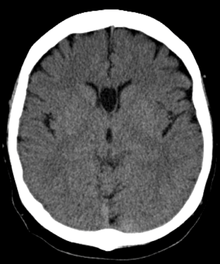
 Stylized horizontal section through the brain showing normal and cave of septum pellucidum. | |
 Difference between cave of septum pellucidum (CSP), cavum vergae (CV), and cavum veli interpositi (CVI). 3=third ventricle, 4=fourth ventricle. | |
| Details | |
| Identifiers | |
| Latin | Cavum septi pellucidi |
| TA | A14.1.09.263 |
| FMA | 61874 |
The cave of septum pellucidum (CSP) describes a septum pellucidum that has a separation between its two leaflets (septal laminae). This cavity contains cerebrospinal fluid (CSF) that filters from the ventricles through the septal laminae.[1]
The cave of septum pellucidum is bounded anteriorly by the genu of the corpus callosum; superiorly by the body of the corpus callosum; posteriorly by the anterior limb and pillars of the fornix; inferiorly by the anterior commissure and the rostrum of the corpus callosum; and laterally by the leaflets of the septum pellucidum.[2]
There are individual differences in the degree of CSP; whereas some have complete closure of the cavum, others present with a small degree (4-6mm in the coronal plane) of incomplete closure.[3]
The most common type of CSP is noncommunicating, that is, it does not connect to the brain's ventricular system. Because of this lack of communication, the erstwhile term for CSP, the "fifth ventricle," is not anatomically correct and its use has fallen out of favor in recent years.
CSP is present in 100% of fetuses, but over 85% of them fuse by 3–6 months after birth.[4][5]
Cause
The cause of CSP is basically unknown, although it is thought that prenatal alcohol exposure plays a significant role.[3]
Marker for fetal neural maldevelopment
CSP is a marker for fetal neural maldevelopment. The septum pellucidum is a thin, triangular, vertical membrane separating the anterior horns of the left and right lateral ventricles of the brain. It runs as a sheet from the corpus callosum down to the fornix. During fetal development at approximately the twelfth week of gestation, a space forms between two laminae, which is the CSP. At approximately the twentieth week of gestation, the laminae start to close. This closure ends shortly after birth (3–6 months postnatally). Fusion of the CSP is attributed to rapid development of the alvei of the hippocampus, amygdala, septal nuclei, fornix, corpus callosum and other midline structures. Lack of such limbic development interrupts this posterior-to-anterior fusion, resulting in preservation of the CSP into adulthood.[3]
Association with mental disorders
CSP has been loosely associated with schizophrenia,[6] post-traumatic stress disorder,[7] traumatic brain injury,[8] as well as with antisocial personality disorder.[3] CSP is one of the distinguishing features of individuals displaying symptoms of dementia pugilistica.[9] For the majority of individuals, CSP produces no ill effects.
References in movies
- In Rocky V, Rocky Balboa is notified he has cavum septum pellucidum, especially after his last fight with Ivan Drago. He is forced to retire as a result.
See also
References
- ↑ Oteruelo F (1986). "On the cavum septi pellucidi and the cavum Vergae". Anatomischer Anzeiger. 162 (4): 271–8. PMID 3813041.
- ↑ Born C, Meisenzahl E, Frodl T, Pfluger T, Reiser M, Möller H, Leinsinger G (2004). "The septum pellucidum and its variants. An MRI study". European archives of psychiatry and clinical neuroscience. 254 (5): 295–302. doi:10.1007/s00406-004-0496-z. PMID 15365704.
- 1 2 3 4 Adrian Raine; Lydia Lee; Yaling Yang; Patrick Colletti (2010). "Neurodevelopmental marker for limbic maldevelopment in antisocial personality disorder and psychopathy". BJPsych. The British Journal of Psychiatry. 197: 186–192. doi:10.1192/bjp.bp.110.078485.
- ↑ Farruggia S, Babcock D (1981). "The cavum septi pellucidi: its appearance and incidence with cranial ultrasonography in infancy". Radiology. 139 (1): 147–50. doi:10.1148/radiology.139.1.7208915. PMID 7208915.
- ↑ "Paediatric Imaging: Cavum septum pellucidum". Medcyclopedia.com.
- ↑ Galarza M, Merlo A, Ingratta A, Albanese E, Albanese A (2004). "Cavum septum pellucidum and its increased prevalence in schizophrenia: a neuroembryological classification". The Journal of neuropsychiatry and clinical neurosciences. 16 (1): 41–6. doi:10.1176/appi.neuropsych.16.1.41. PMID 14990758.
- ↑ May F, Chen Q, Gilbertson M, Shenton M, Pitman R (2004). "Cavum septum pellucidum in monozygotic twins discordant for combat exposure: relationship to posttraumatic stress disorder". Biol. Psychiatry. 55 (6): 656–8. doi:10.1016/j.biopsych.2003.09.018. PMC 2794416
 . PMID 15013837.
 - ↑ Zhang L, Ravdin L, Relkin N, Zimmerman R, Jordan B, Lathan W, Uluğ A (2003). "Increased diffusion in the brain of professional boxers: a preclinical sign of traumatic brain injury?". AJNR. American journal of neuroradiology. 24 (1): 52–7. PMID 12533327.
- ↑ Neuropathol Exp Neurol. 2009 Jul;68(7):709-35. doi: 10.1097/NEN.0b013e3181a9d503. Chronic traumatic encephalopathy in athletes: progressive tauopathy after repetitive head injury. McKee AC, Cantu RC, Nowinski CJ, Hedley-Whyte ET, Gavett BE, Budson AE, Santini VE, Lee HS, Kubilus CA, Stern RA.
External links
| Wikimedia Commons has media related to Cave of septum pellucidum. |
- Mypacs scan by Rolando Reyna, Radiologist, Hospital Santo Tomas, Panama. Good image, but text is incorrect and not referenced.