Bird feet and legs
.jpg)
Most birds are digitigrade animals, which means that they walk on their toes, not the entire foot.[2][3][4][5] Some of their lower bones of the foot[3] (distals[6] and most[3][5] of metatarsals[6]) are fused to form tarsometatarsus[5][7] – a third segment of the leg, specific to birds. The upper bones of the foot[3] (proximals) in turn are fused with the tibia to form tibiotarsus,[5][7] as over time the centralia disappeared.[6] The fibula is also reduced.[3][5][7]
The legs are attached to a very strong assembly consisting of the pelvic girdle extensively fused with the uniform spinal bone (also specific to birds) called the synsacrum, built from some of the fused bones.[3][4][7][8]
Bird leg and feet anatomy is very diverse. It reveals many accommodations to perform a wide variety of functions.[1][2][3][4][5][9]
Skeleton

Hindlimbs
Birds are generally digitigrade animals (toe - walkers)[3][4] as reflected in the structure of their leg skeleton. They use only hindlimbs to walk, which is called bipedalism.[2] As mentioned earlier, most bones of the avian foot (except toes) are fused together or with other bones, having over time changed function.
Tarsometatarsus
Some lower bones of the foot are fused to form tarsometatarsus[3][6][7] – a third segment of the leg specific for birds. It consists of merged distals and metatarsals[6] II, III and IV.[3] Metatarsus I remains separated as a base of the first digit.[3][5] Tarsometatarsus is the extended foot area giving the leg an extra lever length.[3]
Tibiotarsus
The upper bones of the foot[3] (proximals) are fused with the tibia to form tibiotarsus,[5][7] while the centralia disappeared.[6] The anterior (frontal) side of the dorsal end of the tibiotarsus (at the knee) contains a protruding enlargement called the cnemial crest.[2]
Patella
At the knee above the cnemial crest, the patella (kneecap[2]) is located.[5] Some species don't have patellas; sometimes it is only a small extension of the cnemial crest. In grebes there exist both a normal patella and an extension of the cnemial crest.[2]
Fibula
The fibula is reduced[2][3] and adheres extensively to the tibia,[7] usually reaching only 2/3 of its length. Only penguins have full length fibula.[5]
Femur
Knee and ankle - confusions
The bird knee joint between the femur and tibia (or rather tibiotarsus) points obviously forwards,[2] but is hidden in feathers. The backward-pointing "heel" (ankle [2][4]) we can see is a joint between the tibiotarsus and tarsometatarsus.[3][5] The joint inside tarsus occurs also in some reptiles.[7] It is worth noting here that the name "thick knee" of the birds in the family Burhinidae is incorrect, because the heel of these birds is large. So ornithologists confused knee and ankle here.[2]
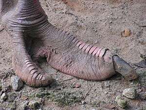
Toes and unfused metatarsals
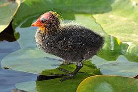
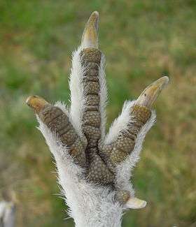
Most birds have 4 toes (digits), typically 3 facing forward and 1 pointing backward.[3][4][7] In a typical perching bird they consist respectively of 3, 4, 5 and 2 phalanges.[2] Some birds have 3 forward-facing toes (tridactyl, for example sanderling[2]) and the ostrich 2 toes (didactyl[2]).[5] The first digit called hallux is homologous to the human big toe[3][4] and usually projects to the rear.[3][4][5]
On the extreme phalanx of each toe, claws[5] are located. They are a horny podotheca or sheath,[2] so are not part of the skeleton.
The bird foot also contains 1 or 2 metatarsals not fused in the tarsometatarsus.[7]
Pelvic girdle and synsacrum
The legs are attached to a very strong (but lightweight[4]) assembly consisting of the pelvic girdle extensively fused with uniform spinal bone called synsacrum,[3][4][7][8] which is specific to birds. Synsacrum is built from fused lumbar, sacral, some of first caudal[7] and sometimes 1–2 last thoracic vertebrae,[8] depending on species - altogether 10–22 vertebrae.[8] Except for ostriches and rheas, pubic bones in the pelvic girdle do not connect each other, making egg-laying easier.[7]
Rigidity and reduction of mass
Fusions of individual bones into rigid strong structures are very characteristic for the bird skeleton.[1][3][4]
Most major bird bones are also extensively pneumatized. They contain many air pockets[2] connected to the pulmonary air sacs[10] of the respiratory system. Their spongelike interior makes them very strong relatively to their mass.[2][3] The number of pneumatic bones however depends on species. It is worth noting that the pneumaticity is slight[11] or absent in diving birds. For example, in the long-tailed duck, the bones of the leg (and wing) are not pneumatic in contrast with some of the other bones, while loons[12] and puffins have even more massive skeletons without any aired bones.[13] Surprisingly, the flightless ostrich and emu have pneumatic femurs and so far this is the only known aired bone in these birds[14] except cervical vertebrae of the ostrich.[10]
Fusions (rigidity)[1] and pneumatic bones (reduction of mass) are considered as some of the many adaptations of birds for flight.[3]
Plantigrade locomotion in birds
Most birds except loons and grebes are digitigrade, not plantigrade.[2] Also chicks in the nest[5] can use the entire foot (toes and tarsometatarsus) with the heel on the ground.
Loons tend to walk this way because their legs and pelvis are deeply specialized for swimming. They have a narrow pelvis, which moves the attachment point of the femur to the rear, and their tibiotarsus is much longer than the femur. This shifts the feet (toes) behind the center of mass of the loon body. They walk usually by pushing themselves on their breasts; larger loons cannot take off from land.[4] This position however is highly suitable for swimming, because their feet are located at the rear like the propeller on a motorboat.[2]
Grebes and many other waterfowl have shorter femur and more or less narrow pelvis too, which gives the impression that their legs are attached to the rear like in loons.[2]
Functions of bird legs and feet

_-feet.jpg)

Because avian forelimbs are converted to wings, many of their functions are performed by the bill and hindlimbs (feet and legs).[4] It has been proposed that the hindlimbs are also very important in flight as accelerators when taking-off.[15][16] Some leg and foot functions, including conventional ones and those specific to birds are:
- locomotion[2][4]
- perching on a branch[2][4] or clinging[4]
- carrying (like osprey holding fish)[2][4]
- flight - related
- feeding - related
- catching[3][4] and killing[2][3] prey in raptors (hawk, owl[2])
- holding[3][4] (parrots - used like hands[2]) and pulling apart[3] food (along with the bill[3])
- scratching the ground in search of food[2]
- "double scratch" - hopping forward and then backward using both feet to scratch (often towhee, sparrow, junco)[2]
- one-footed scratch (grouse, quail, wild turkey, domestic chicken)[2]
- reproduction - related
- cradling [4] and turning[2] eggs during incubation. Birds lacking a brood patch incubate the eggs with their feet - grasping one or even two of them (gannets, boobies) or keeping them on the top surfaces of their feet (penguins under a pouch of belly skin, murres).[1]
- courtship[4] (sage grouse[2]), aerial courtship[4] (bald eagle[2])
- building nests (in addition to the bill)[1]
- preening[2][4] and cleaning.[2] Sometimes birds use a special claw, for example barn owls have a so-called "feather comb", some herons and nightjars use the claw for cleaning the head.[2]
- heat loss regulation (herons, gulls, giant petrel, storks, New World vultures,[1] ducks, geese[2])
Toe arrangements
_EN.gif)
Typical toe arrangements in birds are:
- zygodactyl - two toes in front (2, 3) and two in back (1, 4) - the outermost front toe (4) is reversed[1][4]
- Many perching birds - most woodpeckers[2][4] and their allies,[1] osprey,[4] owls,[4] cuckoos,[4] most parrots,[4][5] mousebirds, some swifts,[1][3] cuckoo roller.[1]
- Woodpeckers when climbing can rotate outer rear digit (4) to the side in an ectropodactyl arrangement. Black-backed woodpecker, Eurasian three-toed woodpecker and American three-toed woodpecker have three toes - the inner rear (1) is missing, outer rear (4) points always backward (never rotates).[4]
- Owls, osprey and turacos can rotate outer toe (4) back and forth.[4]
- The zygodactyl arrangement is a case of convergence, because it evolved in birds in different ways nine times.[1]
- heterodactyl - two toes in front (3, 4) and two in back (2, 1) - the inner front toe (2) is reversed.
- syndactyl - three toes in front (2, 3, 4), one in back (1), two - outer and middle (2, 3) are joined for much of their length.[1][2]
- Common in Coraciiformes,[1] including kingfishers[4] and hornbills.[3]
- pamprodactyl - two inner toes in front (2, 3), two outer (1, 4) can rotate freely forward and backward.[1][2]
- Mousebirds, some swifts.[1][2][3][4]
- Some swifts move all four digits forward to use them as hooks to hang.[3]
Most common is the anisodactyl foot,[2][4][9] second among perching birds is the zygodactyl arrangement.[3]
Webbing and lobation

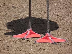
Palmations and lobes enable swimming or help walking on loose ground like over mud.[2][4] The webbed or palmated feet of birds can be categorized into the following types:
- palmate - only the anterior digits (2-4) joined by the webbing.[4]
- Ducks,[1][9] geese[9] and swans,[4] gulls and terns,[4] and other aquatic birds (auks, flamingos, fulmars, jaegers, loons, petrels, shearwaters, skimmers[2]).[2][3]
- Diving ducks additionally have lobed hind toe (1),[2][4] gulls, terns and allies have reduced hind toe.[4]
- Gannets and boobies, pelicans, cormorants,[3][4][9] anhingas, frigatebirds.[2]
- Some gannets have brightly colored feet used in display.[4]
- Some plovers[3][4] (Eurasian dotterel[2]) and sandpipers[3][4] (semipalmated sandpiper, stilt sandpiper, upland sandpiper, greater yellowlegs, willet[2]), avocet, herons (only two toes),[2] all grouse, some domestic breeds of chicken.[3]
- Plovers and lapwings have a vestigial hind toe (1),[4] sandpipers and their allies a reduced and raised hind toe almost not touching the ground.[4] Sanderling is the only sandpipper having 3 toes (tridactyl foot).[2]
- Lobes expand or contract when bird swims.[3]
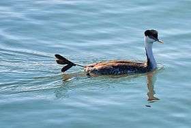
- Grebes,[3][5] coots,[1][5] phalaropes,[4][9] finfoots,[2] some palmate-footed ducks[3] on the hallux (1).[4]
- Grebes have more webbing between the toes in comparison to coots and phalaropes.[4]
Most common is the palmate foot.
Heat loss regulation
Some of birds like gulls, herons,[1] ducks or geese[2] can regulate heat loss by their feet.
The arteries (carrying blood from the heart) and veins (carrying blood toward the heart) intertwine in the legs so heat can be transferred back from arteries to veins before reaching feet. Such a mechanism is called countercurrent exchange. Gulls can also open a shunt between these vessels, turning back the blood stream above the foot, and constrict the vessels in the foot. This reduces heat loss by more than 90 percent. In gulls the temperature of the base of the leg is 32°C (89°F), of the foot may be close to 0°C[1] (32°F).
However, for cooling, this heat-exchange network can be bypassed and blood flow through the foot significantly increased (giant petrel). Some birds additionally excrete onto their feet, increasing heat loss thanks to evaporation (storks, New World vultures).[1]
See also
References
| Wikimedia Commons has media related to Bird feet. |
- 1 2 3 4 5 6 7 8 9 10 11 12 13 14 15 16 17 18 19 20 21 22 23 24 25 26 27 28 Gill, Frank B. (2001). Ornithology (2d ed.). New York: W.H. Freeman and Company. ISBN 0-7167-2415-4.
- 1 2 3 4 5 6 7 8 9 10 11 12 13 14 15 16 17 18 19 20 21 22 23 24 25 26 27 28 29 30 31 32 33 34 35 36 37 38 39 40 41 42 43 44 45 46 47 48 49 50 51 52 53 54 55 56 57 58 59 60 61 62 63 64 65 66 67 68 69 70 71 Kochan, Jack B. (1994). Feet & Legs. Birds. Mechanicsburg: Stackpole Books. ISBN 0-8117-2515-4.
- 1 2 3 4 5 6 7 8 9 10 11 12 13 14 15 16 17 18 19 20 21 22 23 24 25 26 27 28 29 30 31 32 33 34 35 36 37 38 39 40 41 42 43 44 45 46 47 48 49 50 51 Proctor, Noble S.; Lynch, Patrick J. (1993). "Chapters: 6.Topography of the foot; 11.The pelvic gridle; 12.The bones of the leg and foot Family". Manual of Ornithology. Avian Structure & Function. New Haven and London: Yale University Press. pp. 70–75, 140–141, 142–144. ISBN 0-300-07619-3. External link in
|publisher=(help) - 1 2 3 4 5 6 7 8 9 10 11 12 13 14 15 16 17 18 19 20 21 22 23 24 25 26 27 28 29 30 31 32 33 34 35 36 37 38 39 40 41 42 43 44 45 46 47 48 49 50 51 52 53 54 55 56 57 58 59 60 Elphick, John B.; Dunning, JR., Jack B.; Sibley, David Allen (2001). National Audubon Society. The Sibley Guide to Bird Life & Behavior. New York: Alfred A. Knopf. ISBN 0-679-45123-4.
- 1 2 3 4 5 6 7 8 9 10 11 12 13 14 15 16 17 18 19 Kowalska-Dyrcz, Alina (1990). "Entry: noga [leg]". In Busse, Przemysław. Ptaki [Birds]. Mały słownik zoologiczny [Small zoological dictionary] (in Polish). I (I ed.). Warsaw: Wiedza Powszechna. pp. 383–385. ISBN 83-214-0563-0.
- 1 2 3 4 5 6 Romer, Alfred Sherwood; Parsons, Thomas S. (1977). The Vertebrate Body. Philadelphia, PA: Holt-Saunders International. pp. 205–208. ISBN 0-03-910284-X.
- 1 2 3 4 5 6 7 8 9 10 11 12 13 Dobrowolski, Kazimierz A.; Klimaszewski, Sędzimir M.; Szelęgiewicz, Henryk (1981). "Chapters: Gromada: Ptaki - Aves: Układ kostny; Pas miednicowy i kończyna tylna [Class: Birds: The skeletal system; The pelvic girdle and the hindlimb]". Zoologia [Zoology] (in Polish) (4th ed.). Warsaw: Wydawnictwo Szkolne i Pedagogiczne. pp. 462–464, 469. ISBN 83-02-00608-4.
- 1 2 3 4 Kowalska-Dyrcz, Alina (1990). "Entry: synsakrum [synsacrum]". In Busse, Przemysław. Ptaki [Birds]. Mały słownik zoologiczny [Small zoological dictionary] (in Polish). II (I ed.). Warsaw: Wiedza Powszechna. p. 245. ISBN 83-214-0563-0.
- 1 2 3 4 5 6 Kalbe, Lothar (1983). "Besondere Formen für spezielle Aufgaben der Wassertiere [Special adaptations of aquatic animals to specific lifestyles]". Tierwelt am Wasser [Wildlife by the water] (in German) (1st ed.). Leipzig-Jena-Berlin: Urania-Verlag. pp. 72–77.
- 1 2 Wedel, Mathew J. (2003). "Vertebral pneumaticity, air sacs, and the physiology of sauropod dinosaurs" (PDF). Paleobiology. The Paleontological Society. 29 (2): 243–255. doi:10.1666/0094-8373(2003)029<0243:vpasat>2.0.co;2. Retrieved 2014-01-21. External link in
|publisher=(help) - ↑ Schorger, A. W. (September 1947). "The deep diving of the loon and old-squaw and its mechanism" (PDF). The Wilson Bulletin. The Wilson Ornithological Society. 59 (3): 151–159. Retrieved 2014-01-21. External link in
|publisher=(help) - ↑ Gier, H. T. (1952). "The air sacs of the loon" (PDF). The Auk. American Ornithologists' Union. 69 (1): 40–49. doi:10.2307/4081291. Retrieved 21 January 2014.
- ↑ Fastovsky, David E.; Weishampel, David B. (2005). The Evolution and Extinction of the Dinosaurs (second ed.). Cambridge, New York, Melbourne,Madrid, Cape Town, Singapore, São Paulo: Cambridge University Press. ISBN 0-521-81172-4. Retrieved 2014-01-21.
- ↑ Bezuidenhout, A.J.; Groenewald, H.B.; Soley, J.T. (1999). "An anatomical study of the respiratory air sacs in ostriches" (PDF). [www.ojvr.org/ Onderstepoort Journal of Veterinary Research]. The Onderstepoort Veterinary Institute. 66: 317–325. Retrieved 2015-05-22.
- 1 2 3 Earls, Kathleen D. (Feb 2000). "Kinematics and mechanics of ground take-off in the starling Sturnis vulgaris and the quail Coturnix coturnix". The Journal of Experimental Biology. 203: 725–739. PMID 10648214. Retrieved 2014-01-17.
- 1 2 Whitfield, John (10 March 2000). "Off to a flying jump-start : Nature News". Nature Publishing Group, a division of Macmillan Publishers Limited. doi:10.1038/news000316-1. Retrieved 2014-01-17.