B-cell receptor
The B-cell receptor or BCR is a transmembrane receptor protein located on the outer surface of B cells. The receptor's binding moiety is composed of a membrane-bound antibody that, like all antibodies, has a unique and randomly determined antigen-binding site (see V(D)J recombination).[1] When a B cell is activated by its first encounter with an antigen that binds to its receptor (its "cognate antigen"), the cell proliferates and differentiates to generate a population of antibody-secreting plasma B cells and memory B cells. The B cell receptor (BCR) has two crucial functions upon interaction with the antigen. One function is signal transduction, involving changes in receptor oligomerization. The second function is to mediate internalization for subsequent processing of the antigen and presentation of peptides to helper T cells.[1] BCR functions are required for normal antibody production, and defects in BCR signal transduction may lead to immunodeficency, and B-cell malignancy.[1]
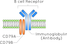
Components of the B-cell receptor
The B-cell receptor is composed of two parts:[1] i) A membrane-bound immunoglobulin molecule of one isotype (IgD, IgM, IgA, IgG, or IgE). With the exception of the presence of an integral membrane domain, these are identical to their secreted forms. ii) Signal transduction moiety: A heterodimer called Ig-α/Ig-β (CD79), bound together by disulfide bridges. Each member of the dimer spans the plasma membrane and has a cytoplasmic tail bearing an immunoreceptor tyrosine-based activation motif (ITAM).
Signaling pathways of the B-cell receptor
There are several signaling pathways that the B-cell receptor can follow through. The physiology of B cells is intimately connected with the function of their B-cell receptor.[1] In B cells, the balance of initiation, amplitude and duration of BCR activation can be influenced by a specific immunoglobulin structure, the expression adaptor molecules (like GAB1, BLNK, GRB2, CARD11), the activity of kinases (like LYN, SYK, PI3K) or phosphatases (like SHIP-1, SHP-1 and PTEN) and levels of microRNAs. [2] [3] The miR-150 and miR-155 were shown to be regulators of proteins that affect the propensity of BCR signalling pathway.[4][5]
- IKK/NF-κB Transcription Factor Pathway: CD79 and other proteins, microsignalosomes, go to activate PLC-γ after antigen recognition by the BCR and before it goes to associate into the c-SMAC. It then cleaves PIP2 into IP3 and DAG (diacylglycerol). IP3 acts as a second messenger to dramatically increase ionic calcium inside the cytosol (via release from the endoplasmic reticulum or influx from the extracellular environment via ion channels). This leads to eventual activation of PKCβ from the calcium and DAG. PKCβ phosphorylates (either directly or indirectly) the NF-κB signaling complex protein CARMA1 (the complex itself comprising CARMA1, BCL10, and MALT1). These result in recruitment and summoning of the IKK (IkB kinase), TAK1, by several ubiquitylation enzymes also associated with the CARMA1/BCL10/MALT1 complex. MALT1 itself is a caspase-like protein that cleaves A20, an inhibitory protein of NF-κB signaling (which acts by deubiquitylating NF-κB’s ubiquitylation substrates, having an inhibitory effect). TAK1 phosphorylates the IKK trimer after it too has been recruited to the signaling complex by its associated ubiquitylation enzymes. IKK then phosphorylates IkB (an inhibitor of and bound to NF-κB), which induces its destruction by marking it for proteolytic degradation, freeing cytosolic NF-κB. NF-κB then migrates to the nucleus to bind to DNA at specific response elements, causing recruitment of transcription molecules and beginning the transcription process.[1]
2. Ligand binding to the BCR also leads to the phosphorylation of the protein BCAP. This leads to the binding and activation of several proteins with phosphotyrosine-binding SH2 domains. One of these proteins is PI3K. Activation of PI3K leads to PIP2 phosphorylation, forming PIP3. Proteins with PH (Pleckstrin homology) domains can bind to the newly created PIP3 and become activated. These include proteins of the FoxO family, which stimulate cell cycle progression, and protein kinase D, which enhances glucose metabolism. Another important protein with a PH domain is Bam32. This recruits and activates small GTPases such as Rac1 and Cdc42. These, in turn, are responsible for the cytoskeletal changes associated with BCR activation by modifying actin polymerisation.
The B-cell receptor in malignancy
The B-cell receptor has been shown to be involved in the pathogenesis of various B cell-derived lymphoid cancers.[1][3] Although it may be possible that stimulation by antigen binding contributes to the proliferation of malignant B cells,[6] increasing evidence implicates antigen-independent self-association of BCRs as a key feature in a growing number of B-cell neoplasias.[1][7][8][9][10] B-cell receptor signalling is currently a therapeutic target in various lymphoid neoplasms.
References
- 1 2 3 4 5 6 7 8 Seda, V; Mraz, M (2014). "B cell receptor (BCR) signalling and its crosstalk with other pathways in normal and malignant cells". European Journal of Haematology. 94: n/a. doi:10.1111/ejh.12427. PMID 25080849.
- ↑ Packard, T.A.; Cambier, J.C. (2013). "B lymphocyte antigen receptor signaling: initiation, amplification, and regulation.". F1000Prime Rep. 5 (40). doi:10.12703/P5-40. PMC 3790562
 . PMID 24167721.
 - 1 2 Mraz, M.; Kipps, T. J. (2013). "MicroRNAs and B Cell Receptor Signaling in Chronic Lymphocytic Leukemia". Leukemia & Lymphoma. 54 (8): 1836–9. doi:10.3109/10428194.2013.796055. PMID 23597135.
- ↑ "MicroRNA-155 influences B-cell receptor signaling and associates with aggressive disease in chronic lymphocytic leukemia". Blood. 124: 546–54. 2014. doi:10.1182/blood-2014-03-559690. PMC 4110661. PMID 24914134.
- ↑ "miR-150 influences B-cell receptor signaling in chronic lymphocytic leukemia by regulating expression of GAB1 and FOXP1.". Blood. 124: 84–95. Jul 2014. doi:10.1182/blood-2013-09-527234. PMID 24787006.
- ↑ Daneshek, W; RS Schwartz (1959). "Leukemia and auto-immunization- some possible relationships". Blood. 14.
- ↑ Corcos, D (1990). "Oncogenic potential of the B-cell antigen receptor and its relevance to heavy chain diseases and other B-cell neoplasias: a new model". Res Immunol. 141 (6): 543–53. doi:10.1016/0923-2494(90)90022-Q. PMID 2284498.
- ↑ Corcos D, Dunda O, Butor C, et al. (1995). "Pre-B-cell development in the absence of lambda 5 in transgenic mice expressing a heavy-chain disease protein". Curr. Biol. 5 (10): 1140–8. doi:10.1016/S0960-9822(95)00230-2. PMID 8548286.
- ↑ Davis, RE; Ngo, Vu N.; Lenz, G; Tolar, P; Young, RM; Romesser, PB; Kohlhammer, H; Lamy, L; Zhao, H; Yang, Y; Xu, W; Shaffer, AL; Wright, G; Xiao, W; Powell, J; Jiang, JK; Thomas, CJ; Rosenwald, A; Ott, G; Muller-Hermelink, HK; Gascoyne, RD; Connors, JM; Johnson, NA; Rimsza, LM; Campo, E; Jaffe, ES; Wilson, WH; Delabie, J; Smeland, EB; Fisher, RI; Braziel, RM; Tubbs, RR; Cook, JR; Weisenburger, DD; Chan, WC; Pierce, SK; Staudt, LM (2010). "Chronic active B-cell-receptor signalling in diffuse large B-cell lymphoma". Nature. 463 (7277): 88–92. Bibcode:2010Natur.463...88D. doi:10.1038/nature08638. PMC 2845535. PMID 20054396.
- ↑ Dühren-von Minden M; Übelhart R; Schneider D; Wossning T; Bach MP; Buchner M; Hofmann D; Surova E; Follo M; Köhler F; Wardemann H; Zirlik K; Veelken H; Jumaa H (2012). "Chronic lymphocytic leukaemia is driven by antigen-independent cell-autonomous signalling". Nature. 489 (7415): 309–312. Bibcode:2012Natur.489..309M. doi:10.1038/nature11309. PMID 22885698.
External links
- B-Cell Antigen Receptors at the US National Library of Medicine Medical Subject Headings (MeSH)