ATP synthase, H+ transporting, mitochondrial F1 complex, alpha 1
| View/Edit Human | View/Edit Mouse |
ATP synthase subunit alpha, mitochondrial is an enzyme that in humans is encoded by the ATP5A1 gene.[3][4]
This gene encodes a subunit of mitochondrial ATP synthase. Mitochondrial ATP synthase catalyzes ATP synthesis, using an electrochemical gradient of protons across the inner membrane during oxidative phosphorylation. ATP synthase is composed of two linked multi-subunit complexes: the soluble catalytic core, F1, and the membrane-spanning component, Fo, comprising the proton channel. The catalytic portion of mitochondrial ATP synthase consists of 5 different subunits (alpha, beta, gamma, delta, and epsilon) assembled with a stoichiometry of 3 alpha, 3 beta, and a single representative of the other 3. The proton channel consists of three main subunits (a, b, c). This gene encodes the alpha subunit of the catalytic core. Alternatively spliced transcript variants encoding the same protein have been identified. Pseudogenes of this gene are located on chromosomes 9, 2, and 16.[4]
Structure
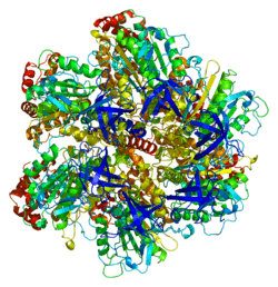
The ATP5A1 gene, located on the q arm of chromosome 18 in position 21, is made up of 13 exons and is 20,090 base pairs in length.[4] The ATP5A1 protein weighs 59.7 kDa and is composed of 553 amino acids.[5][6] The protein is a subunit of the catalytic portion of the F1Fo ATPase, also known as Complex V, which consists of 14 nuclear and 2 mitochondrial -encoded subunits. As an alpha subunit, ATP5A1 is contained within the catalytic F1 portion of the complex.[4] The nomenclature of the enzyme has a long history. The F1 fraction derives its name from the term "Fraction 1" and Fo (written as a subscript letter "o", not "zero") derives its name from being the binding fraction for oligomycin, a type of naturally-derived antibiotic that is able to inhibit the Fo unit of ATP synthase.[7][8] The F1 particle is large and can be seen in the transmission electron microscope by negative staining.[9] These are particles of 9 nm diameter that pepper the inner mitochondrial membrane. They were originally called elementary particles and were thought to contain the entire respiratory apparatus of the mitochondrion, but, through a long series of experiments, Efraim Racker and his colleagues (who first isolated the F1 particle in 1961) were able to show that this particle is correlated with ATPase activity in uncoupled mitochondria and with the ATPase activity in submitochondrial particles created by exposing mitochondria to ultrasound. This ATPase activity was further associated with the creation of ATP by a long series of experiments in many laboratories.
Function
Mitochondrial membrane ATP synthase (F1Fo ATP synthase or Complex V) produces ATP from ADP in the presence of a proton gradient across the membrane which is generated by electron transport complexes of the respiratory chain. F-type ATPases consist of two structural domains, F1 - containing the extramembraneous catalytic core, and Fo - containing the membrane proton channel, linked together by a central stalk and a peripheral stalk. During catalysis, ATP synthesis in the catalytic domain of F1 is coupled via a rotary mechanism of the central stalk subunits to proton translocation. Subunits alpha and beta form the catalytic core in F1. Rotation of the central stalk against the surrounding alpha(3)beta(3) subunits leads to hydrolysis of ATP in three separate catalytic sites on the beta subunits. Subunit alpha does not bear the catalytic high-affinity ATP-binding sites.[10]
Clinical significance
Mutations affecting the ATP5A1 gene cause combined oxidative phosphorylation deficiency 22 (COXPD22), a mitochondrial disorder characterized by intrauterine growth retardation, microcephaly, hypotonia, pulmonary hypertension, failure to thrive, encephalopathy, and heart failure. Mutations on the ATP5A1 gene also cause mitochondrial complex V deficiency, nuclear 4 (MC5DN4), a mitochondrial disorder with heterogeneous clinical manifestations including dysmorphic features, psychomotor retardation, hypotonia, growth retardation, cardiomyopathy, enlarged liver, hypoplastic kidneys and elevated lactate levels in urine, plasma and cerebrospinal fluid.[11]
Model organisms
| Characteristic | Phenotype |
|---|---|
| Homozygote viability | Abnormal |
| Recessive lethal study | Abnormal |
| Body weight | Abnormal[12] |
| Anxiety | Normal |
| Neurological assessment | Normal |
| Grip strength | Normal |
| Hot plate | Normal |
| Dysmorphology | Normal |
| Indirect calorimetry | Normal |
| Glucose tolerance test | Normal |
| Auditory brainstem response | Normal |
| DEXA | Abnormal[13] |
| Radiography | Normal |
| Body temperature | Normal |
| Eye morphology | Normal |
| Clinical chemistry | Abnormal[14] |
| Haematology | Normal |
| Peripheral blood lymphocytes | Normal |
| Micronucleus test | Normal |
| Heart weight | Normal |
| Eye Histopathology | Normal |
| Citrobacter infection | Normal[15] |
| All tests and analysis from[16][17] |
Model organisms have been used in the study of ATP5A1 function. A conditional knockout mouse line, called Atp5a1tm1a(EUCOMM)Wtsi[18][19] was generated as part of the International Knockout Mouse Consortium program — a high-throughput mutagenesis project to generate and distribute animal models of disease to interested scientists.[20][21][22]
Male and female animals underwent a standardized phenotypic screen to determine the effects of deletion.[16][23] Twenty two tests were carried out on mutant mice and five significant abnormalities were observed.[16] No homozygous mutant embryos were identified during gestation, and therefore none survived until weaning. The remaining tests were carried out on heterozygous mutant adult mice and decreased body weight, lean body mass and hypoproteinemia was observed in female animals.[16]
References
- ↑ "Human PubMed Reference:".
- ↑ "Mouse PubMed Reference:".
- ↑ Kataoka H, Biswas C (Sep 1991). "Nucleotide sequence of a cDNA for the alpha subunit of human mitochondrial ATP synthase". Biochim Biophys Acta. 1089 (3): 393–5. doi:10.1016/0167-4781(91)90183-m. PMID 1830491.
- 1 2 3 4 "Entrez Gene: ATP5A1 ATP synthase, H+ transporting, mitochondrial F1 complex, alpha subunit 1, cardiac muscle".
- ↑ Zong NC, Li H, Li H, Lam MP, Jimenez RC, Kim CS, Deng N, Kim AK, Choi JH, Zelaya I, Liem D, Meyer D, Odeberg J, Fang C, Lu HJ, Xu T, Weiss J, Duan H, Uhlen M, Yates JR, Apweiler R, Ge J, Hermjakob H, Ping P (Oct 2013). "Integration of cardiac proteome biology and medicine by a specialized knowledgebase". Circulation Research. 113 (9): 1043–53. doi:10.1161/CIRCRESAHA.113.301151. PMC 4076475
 . PMID 23965338.
 - ↑ "ATP synthase subunit alpha, mitochondrial". Cardiac Organellar Protein Atlas Knowledgebase (COPaKB).
- ↑ Kagawa Y, Racker E (1966). "Partial resolution of the enzymes catalyzing oxidative phosphorylation. 8. Properties of a factor conferring oligomycin sensitivity on mitochondrial adenosine triphosphatase.". Journal of Biological Chemistry. 241: 2461–2466. PMID 4223640.
- ↑ Mccarty RE (November 1992). "A plant biochemist's view of H+
-ATPases and ATP synthases". J. Exp. Biol. 172 (Pt 1): 431–441. PMID 9874753. - ↑ Fernández-Morán H, Oda T, Blair PV, Green DE (July 1964). "A macromolecular repeating unit of mitochondrial structure and function. Correlated electron microscopic and biochemical studies of isolated mitochondria and submitochondrial particles of beef heart muscle". J. Cell Biol. 22 (1): 63–100. doi:10.1083/jcb.22.1.63. PMC 2106494. PMID 14195622.
- ↑ "ATP synthase subunit alpha, mitochondrial". UniProt. The UniProt Consortium.
- ↑ "ATP5A1". NCBI Genetics Home Resource.
- ↑ "Body weight data for Atp5a1". Wellcome Trust Sanger Institute.
- ↑ "DEXA data for Atp5a1". Wellcome Trust Sanger Institute.
- ↑ "Clinical chemistry data for Atp5a1". Wellcome Trust Sanger Institute.
- ↑ "Citrobacter infection data for Atp5a1". Wellcome Trust Sanger Institute.
- 1 2 3 4 Gerdin AK (2010). "The Sanger Mouse Genetics Programme: High throughput characterisation of knockout mice". Acta Ophthalmologica. 88: 925–7. doi:10.1111/j.1755-3768.2010.4142.x.
- ↑ Mouse Resources Portal, Wellcome Trust Sanger Institute.
- ↑ "International Knockout Mouse Consortium".
- ↑ "Mouse Genome Informatics".
- ↑ Skarnes, W. C.; Rosen, B.; West, A. P.; Koutsourakis, M.; Bushell, W.; Iyer, V.; Mujica, A. O.; Thomas, M.; Harrow, J.; Cox, T.; Jackson, D.; Severin, J.; Biggs, P.; Fu, J.; Nefedov, M.; De Jong, P. J.; Stewart, A. F.; Bradley, A. (2011). "A conditional knockout resource for the genome-wide study of mouse gene function". Nature. 474 (7351): 337–342. doi:10.1038/nature10163. PMC 3572410. PMID 21677750.
- ↑ Dolgin E (2011). "Mouse library set to be knockout". Nature. 474 (7351): 262–3. doi:10.1038/474262a. PMID 21677718.
- ↑ Collins FS, Rossant J, Wurst W (2007). "A Mouse for All Reasons". Cell. 128 (1): 9–13. doi:10.1016/j.cell.2006.12.018. PMID 17218247.
- ↑ van der Weyden L, White JK, Adams DJ, Logan DW (2011). "The mouse genetics toolkit: revealing function and mechanism.". Genome Biol. 12 (6): 224. doi:10.1186/gb-2011-12-6-224. PMC 3218837. PMID 21722353.
Further reading
- Dawson SJ, White LA (1992). "Treatment of Haemophilus aphrophilus endocarditis with ciprofloxacin.". J. Infect. 24 (3): 317–20. doi:10.1016/S0163-4453(05)80037-4. PMID 1602151.
- Kovalyov LI, Shishkin SS, Efimochkin AS, et al. (1996). "The major protein expression profile and two-dimensional protein database of human heart.". Electrophoresis. 16 (7): 1160–9. doi:10.1002/elps.11501601192. PMID 7498159.
- Abrahams JP, Leslie AG, Lutter R, Walker JE (1994). "Structure at 2.8 A resolution of F1-ATPase from bovine heart mitochondria.". Nature. 370 (6491): 621–8. doi:10.1038/370621a0. PMID 8065448.
- Akiyama S, Endo H, Inohara N, et al. (1994). "Gene structure and cell type-specific expression of the human ATP synthase alpha subunit.". Biochim. Biophys. Acta. 1219 (1): 129–40. doi:10.1016/0167-4781(94)90255-0. PMID 8086450.
- Jabs EW, Thomas PJ, Bernstein M, et al. (1994). "Chromosomal localization of genes required for the terminal steps of oxidative metabolism: alpha and gamma subunits of ATP synthase and the phosphate carrier.". Hum. Genet. 93 (5): 600–2. doi:10.1007/bf00202832. PMID 8168843.
- Godbout R, Bisgrove DA, Honoré LH, Day RS (1993). "Amplification of the gene encoding the alpha-subunit of the mitochondrial ATP synthase complex in a human retinoblastoma cell line.". Gene. 123 (2): 195–201. doi:10.1016/0378-1119(93)90124-L. PMID 8428659.
- Godbout R, Pandita A, Beatty B, et al. (1997). "Comparative genomic hybridization analysis of Y79 and FISH mapping indicate the amplified human mitochondrial ATP synthase alpha-subunit gene (ATP5A) maps to chromosome 18q12→q21.". Cytogenet. Cell Genet. 77 (3-4): 253–6. doi:10.1159/000134588. PMID 9284928.
- Elston T, Wang H, Oster G (1998). "Energy transduction in ATP synthase.". Nature. 391 (6666): 510–3. doi:10.1038/35185. PMID 9461222.
- Wang H, Oster G (1998). "Energy transduction in the F1 motor of ATP synthase.". Nature. 396 (6708): 279–82. doi:10.1038/24409. PMID 9834036.
- Moser TL, Stack MS, Asplin I, et al. (1999). "Angiostatin binds ATP synthase on the surface of human endothelial cells.". Proc. Natl. Acad. Sci. U.S.A. 96 (6): 2811–6. doi:10.1073/pnas.96.6.2811. PMC 15851. PMID 10077593.
- Moser TL, Kenan DJ, Ashley TA, et al. (2001). "Endothelial cell surface F1-F0 ATP synthase is active in ATP synthesis and is inhibited by angiostatin.". Proc. Natl. Acad. Sci. U.S.A. 98 (12): 6656–61. doi:10.1073/pnas.131067798. PMC 34409. PMID 11381144.
- Wang ZG, White PS, Ackerman SH (2001). "Atp11p and Atp12p are assembly factors for the F(1)-ATPase in human mitochondria.". J. Biol. Chem. 276 (33): 30773–8. doi:10.1074/jbc.M104133200. PMID 11410595.
- Chang SY, Park SG, Kim S, Kang CY (2002). "Interaction of the C-terminal domain of p43 and the alpha subunit of ATP synthase. Its functional implication in endothelial cell proliferation.". J. Biol. Chem. 277 (10): 8388–94. doi:10.1074/jbc.M108792200. PMID 11741979.
- Strausberg RL, Feingold EA, Grouse LH, et al. (2003). "Generation and initial analysis of more than 15,000 full-length human and mouse cDNA sequences.". Proc. Natl. Acad. Sci. U.S.A. 99 (26): 16899–903. doi:10.1073/pnas.242603899. PMC 139241. PMID 12477932.
- Sergeant N, Wattez A, Galván-valencia M, et al. (2003). "Association of ATP synthase alpha-chain with neurofibrillary degeneration in Alzheimer's disease.". Neuroscience. 117 (2): 293–303. doi:10.1016/S0306-4522(02)00747-9. PMID 12614671.
- Ota T, Suzuki Y, Nishikawa T, et al. (2004). "Complete sequencing and characterization of 21,243 full-length human cDNAs.". Nat. Genet. 36 (1): 40–5. doi:10.1038/ng1285. PMID 14702039.
- Bouwmeester T, Bauch A, Ruffner H, et al. (2004). "A physical and functional map of the human TNF-alpha/NF-kappa B signal transduction pathway.". Nat. Cell Biol. 6 (2): 97–105. doi:10.1038/ncb1086. PMID 14743216.
- Cross RL (2004). "Molecular motors: turning the ATP motor.". Nature. 427 (6973): 407–8. doi:10.1038/427407b. PMID 14749816.
- Jin J, Smith FD, Stark C, et al. (2004). "Proteomic, functional, and domain-based analysis of in vivo 14-3-3 binding proteins involved in cytoskeletal regulation and cellular organization.". Curr. Biol. 14 (16): 1436–50. doi:10.1016/j.cub.2004.07.051. PMID 15324660.
PDB gallery | ||||||||||||||||||||||||||||||||||
|---|---|---|---|---|---|---|---|---|---|---|---|---|---|---|---|---|---|---|---|---|---|---|---|---|---|---|---|---|---|---|---|---|---|---|
| ||||||||||||||||||||||||||||||||||


This article incorporates text from the United States National Library of Medicine, which is in the public domain.